Comparison of different segmentation algorithms on brain volume and fractal dimension in infant brain MRIs
Reading time: 5 minute
...
📝 Original Info
Title: Comparison of different segmentation algorithms on brain volume and fractal dimension in infant brain MRIs
ArXiv ID: 2512.12222
Date: 2025-12-13
Authors: - Nathalie Alexander - Arnaud Gucciardi - Umberto Michelucci
📝 Abstract
Accurate segmentation of infant brain MRI is essential for quantifying developmental changes in structure and complexity. However, ongoing myelination and reduced tissue contrast make automated segmentation particularly challenging. This study systematically compared segmentation accuracy and its impact on volumetric and fractal dimension (FD) estimates in infant brain MRI using the Baby Open Brains (BOB) dataset (71 scans, 1-9 months). Two methods, SynthSeg and SamSeg, were evaluated against expert annotations using Dice, Intersection over Union, 95th-percentile Hausdorff distance, and Normalised Mutual Information. SynthSeg outperformed SamSeg across all quality metrics (mean Dice > 0.8 for major regions) and provided volumetric estimates closely matching the manual reference (mean +4% [-28% - 71%]). SamSeg systematically overestimated ventricular and whole-brain volumes (mean +76% [-12% - 190%]). Segmentation accuracy improved with age, consistent with increasing tissue contrast during myelination. Fractal dimension a(FD) nalyses revealed significant regional differences between SynthSeg and expert segmentations, and Bland-Altman limits of agreement indicated that segmentation-related FD variability exceeded most group differences reported in developmental cohorts. Volume and FD deviations were positively correlated across structures, indicating that segmentation bias directly affects FD estimation. Overall, SynthSeg provided the most reliable volumetric and FD results for paediatric MRI, yet small morphological differences in volume and FD should be interpreted with caution due to segmentation-related uncertainty.
💡 Deep Analysis
📄 Full Content
Graphical Abstract
Comparison of different segmentation algorithms on brain volume
and fractal dimension in infant brain MRIs
Nathalie Alexander, Arnaud Gucciardi, Umberto Michelucci
arXiv:2512.12222v1 [cs.CV] 13 Dec 2025
Highlights
Comparison of different segmentation algorithms on brain volume
and fractal dimension in infant brain MRIs
Nathalie Alexander, Arnaud Gucciardi, Umberto Michelucci
• SynthSeg yields more accurate infant brain MRI segmentations than
SamSeg
• Segmentation reliability increases with age and ongoing myelination
• Segmentation bias jointly distorts regional brain volume and fractal
dimension
• Segmentation uncertainty can exceed reported developmental fractal
dimension effects
Comparison of different segmentation algorithms on
brain volume and fractal dimension in infant brain MRIs
Nathalie Alexandera,b, Arnaud Gucciardic,d, Umberto Micheluccic,b
aLaboratory for Motion Analysis, Devision of Paediatric Orthopaedic, Children’s Hospital
of Eastern Switzerland, St. Gallen, Switzerland
bLucerne University of Applied Sciences and Arts, Lucerne, Switzerland
cTOELT llc, Machine Learning Research and Development, Dübendorf, Switzerland
dUniversity of Ljubljana, Faculty of Computer and Information
Science, Ljubljana, Slovenia
Abstract
Accurate segmentation of infant brain MRI is essential for quantifying devel-
opmental changes in structure and complexity. However, ongoing myelination
and reduced tissue contrast make automated segmentation particularly chal-
lenging. This study systematically compared segmentation accuracy and its
impact on volumetric and fractal dimension (FD) estimates in infant brain
MRI using the Baby Open Brains (BOB) dataset (71 scans, 19 months). Two
methods, SynthSeg and SamSeg, were evaluated against expert annotations
using Dice, Intersection over Union, 95th-percentile Hausdorffdistance, and
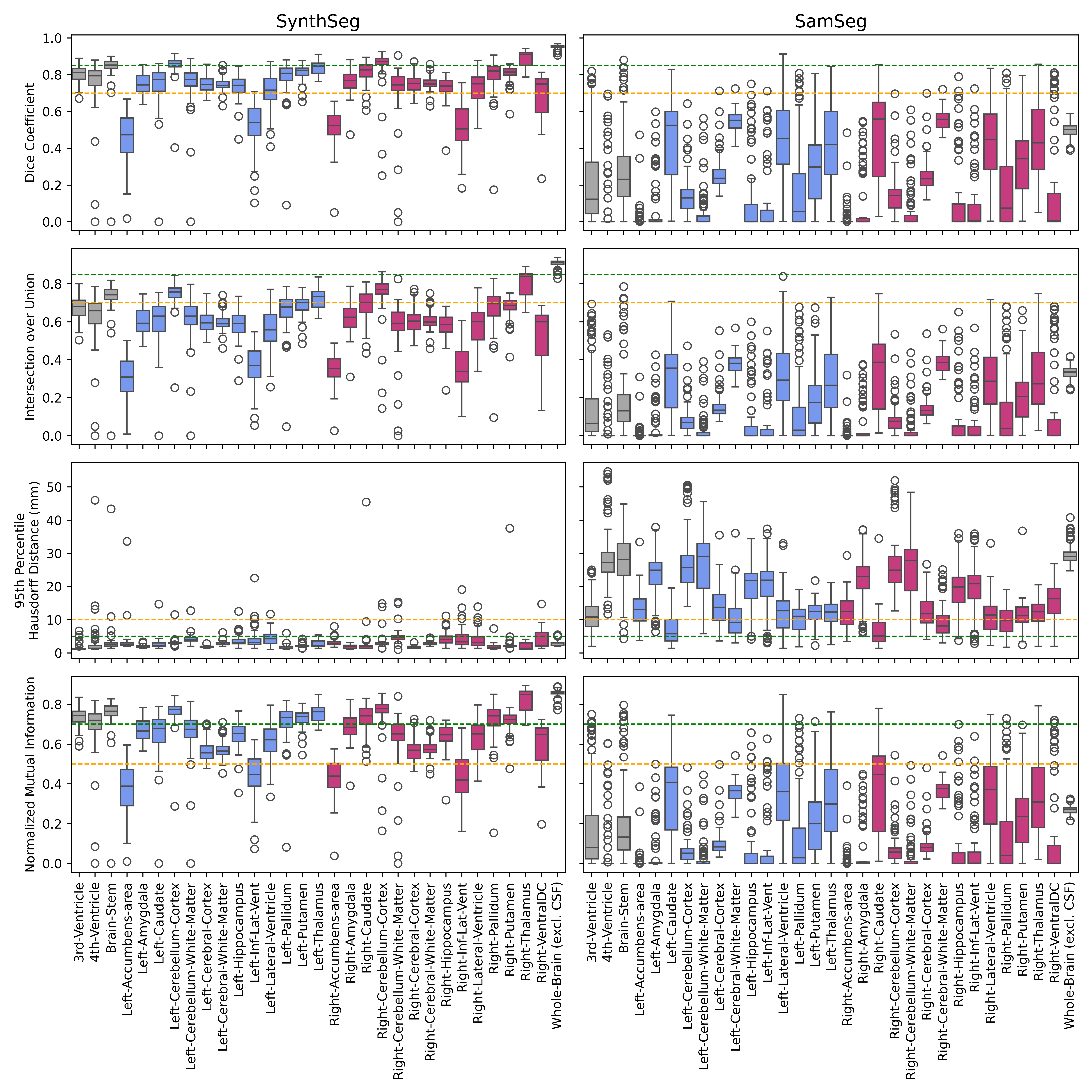
Normalised Mutual Information. SynthSeg outperformed SamSeg across all
quality metrics (mean Dice > 0.8 for major regions) and provided volumetric
estimates closely matching the manual reference (mean +4% [-28% - 71%]).
SamSeg systematically overestimated ventricular and whole-brain volumes
(mean +76% [-12% - 190%]).
Segmentation accuracy improved with age,
consistent with increasing tissue contrast during myelination. FD analyses
revealed significant regional differences between SynthSeg and expert segmen-
tations, and BlandAltman limits of agreement indicated that segmentation-
related FD variability exceeded most group differences reported in develop-
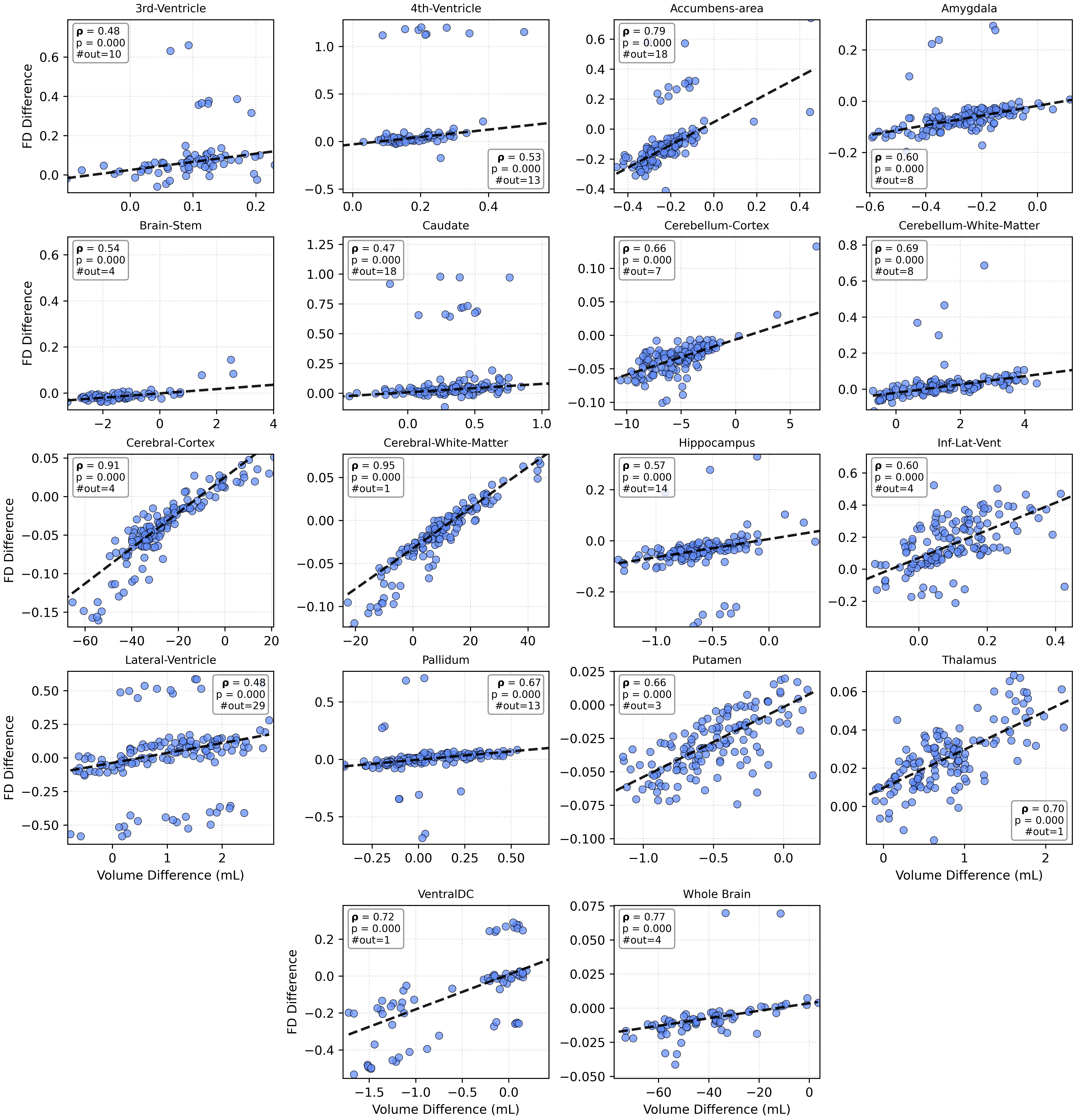
mental cohorts. Volume and FD deviations were positively correlated across
structures, indicating that segmentation bias directly affects FD estimation.
Overall, SynthSeg provided the most reliable volumetric and FD results for
paediatric MRI, yet small morphological differences in volume and FD should
be interpreted with caution due to segmentation-related uncertainty.
Keywords:
Fractal Dimension, Neonatal, Segmentation, Pediatric
1. Introduction
The human brain is a complex organ, with several regions [1]. Magnetic
resonance imaging (MRI) is widely used to investigate brain structures and
tissue composition in vivo. The T1-weighted sequences provide high anatom-
ical detail, with cerebrospinal fluid (CSF) appearing dark and fat bright,
making them suitable for structural evaluation. In contrast, T2-weighted
imaging highlights pathology, as the CSF appears bright and lesions such as
edoema or demyelination are more noticeable due to longer T2 relaxation
times [2].
Within these modalities, brain segmentation is a critical step
that forms the basis for subsequent analyses such as cortical surface recon-
struction and volumetric quantification. However, processing infant brain
magnetic resonance images is particularly challenging compared to adults,
as ongoing myelination leads to reduced tissue contrast, age-dependent in-
tensity changes, and regionally heterogeneous appearances, compounded by
small brain size and partial volume effects. Myelination begins before birth
and continues to increase throughout the first three years of life [3, 4]. A
recent review [5] suggests that myelination is an ongoing process throughout
life and that myelin changes can be divided into five different stages during
human life: early childhood, childhood, adolescence, adult myelination, and
age-related decline in myelin.
Brain development in childhood is characterised by dynamic and region-
specific volumetric changes. The total volume of the brain reaches about
95% of adult size at age six [6, 7], with grey matter following an inverted
U-shaped trajectory and white matter continuing to increase until early
adulthood due to myelination [7]. Individual structures show distinct pat-
terns, such as non-linear hippocampal growth [8] or age-related ventricular
enlargement, which is also diagnostically relevant in hydrocephalus [9, 10].
Altered volumes have been reported across neurodevelopmental conditions,
including enlarged brains in autism [11], reduced cerebral and