Multiple Myeloma (MM) is a blood cancer implying bone marrow involvement, renal damages and osteolytic lesions. The skeleton involvement of MM is at the core of the present paper, exploiting radiomics and artificial intelligence to identify image-based biomarkers for MM. Preliminary results show that MM is associated to an extension of the intrabone volume for the whole body and that machine learning can identify CT image features mostly correlating with the disease evolution. This computational approach allows an automatic stratification of MM patients relying of these biomarkers and the formulation of a prognostic procedure for determining the disease follow-up.
Multiple Myeloma (MM) is a blood cancer implying bone marrow involvement, renal damages and osteolytic lesions. Skeleton involvement of MM is at the core of the present paper, which introduces artificial intelligence and radiomics approaches for the identification of image-based biomarkers for MM. CT data already allows early identification of MM progression and the assessment of the positive response to chemotherapy. However, CT potentials in this field are far to be fully exploited; in particular, there is currently no image-based prognostic index predicting the evolvement of MM. We investigate whether this kind of indices can be identified in either the volumetric dimension of the intrabone space for the whole skeleton asset, or in the imaging features extracted from the focal lesion.
Image processing tools for the analysis of FDG-PET/CT data have been recently developed quantitatively assessing the composition of the skeleton asset and of FDG metabolism in bone marrow. These algorithms utilize pattern recognition in whole-body CT images of patients to segment the compact bone tissue from the bone marrow hosting intrabone volume. In this way, a normalcy database has been constructed [1], which has been utilized to identify the intra-bone volume as a prognostic marker for chronic lymphatic leukemia (CLL) [2] and to associate such alterations to functional modifications in the bone marrow asset of allogeneic transplant [3]. Even more recently, machine learning methods have been applied to features extracted from different imaging modalities within the framework of radiomics paradigms for disease assessment. A few of these methods perform the evaluation of bone formation, regeneration, and asset and, in some cases, these algorithms are able to identify image features that mostly impact the prediction of the disease follow-up. However, none of these methods have been applied so far to CT data from MM patients and therefore the purpose of this paper is two-fold:
• To verify that, as in the case of CLL, intrabone volume represents a reliable biomarker for determining the evolution of MM.
• To verify that the image properties of the compact bone focal lesions can be used for patients’ stratification.
The plan of the paper is as follows. Section 2 illustrates the data used for the analysis and the computational methods for their processing. Section 3 describes the results obtained from the data analysis. Section 4 provides some comments about these results. Our conclusions are offered in Section 5.
The study was performed in accordance with the current version of the Declaration of Helsinki and the International Conference on Harmonization of Good Clinical Practice Guidelines. All patients signed a written informed consent form, encompassing the use of anonimized data for retrospective research purposes, before CT examination. Radiomic analysis was applied to CT data collected in the clinical workup and did not influence patient care in any way.
As far as the design of this retrospective study and the corresponding inclusion criteria are concerned, we have considered 25 consecutive patients (mean age, 62 years ± 8.3; range, 35-70 years) admitted to the IRCCS Policlinico San Martino Hospital because they were suspected of having MM in the last five years. Inclusion criteria were baseline whole-body CT available and retrievable from the Hospital PACS or available from outpatient clinic. The imaging technical standards are minimal and reported in Table 1.
Data obtained in patients’ population were compared to data from 102 control subjects with no history of hematological disease, selected from a previously published normalcy database [1].
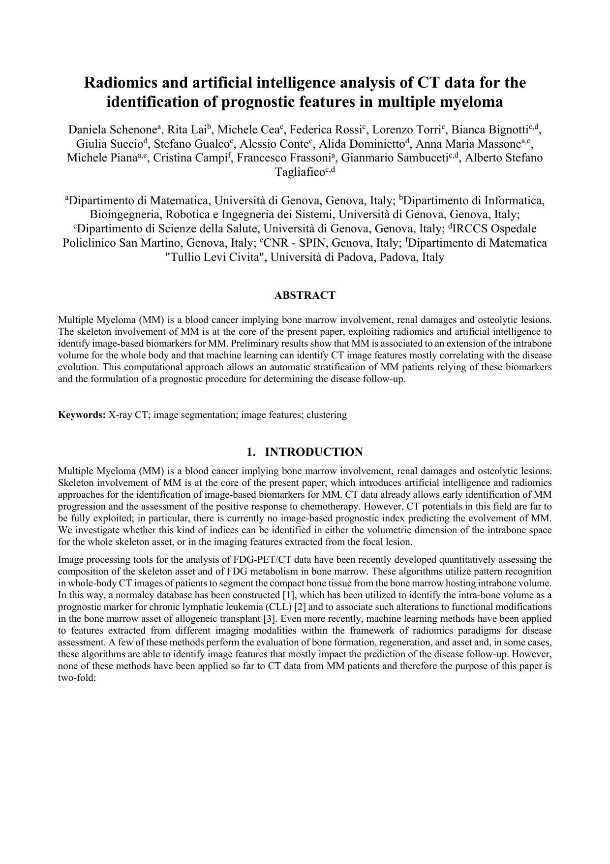
First, the global CT information was processed in order to determine the intraosseous volume potentially available for bone marrow. In order to determine this intrabone volume (IBV) we utilized an already published software code whose underlying assumption is that the Hounsfield value is highest in compact bone among all tissues. Given the fact that attenuation coefficients differ in bones belonging to different districts, we could not apply a mere thresholding technique and therefore we relied on a pattern recognition process based on active contours (see Figure 1). More precisely, the process starts with the unique human intervention asking the operator to draw a loose region of interest around the skull vertex as a starting reference. Then, the functional representing the energy of a curve surrounding the bone profile is iteratively minimized thus determining a sequence of active contours that progressively adapt themselves onto the compact bone border. This procedure is automatically replicated to all slices. In particular, the optimized active contour obtained at the end of the processing of the first slice is utilized as initialization contour for the successive slice. The final 3D result is thus displayed to the operator for the removal of not-bone calcified region
This content is AI-processed based on open access ArXiv data.