The results of chest X-ray (CXR) analysis of 2D images to get the statistically reliable predictions (availability of tuberculosis) by computer-aided diagnosis (CADx) on the basis of deep learning are presented. They demonstrate the efficiency of lung segmentation, lossless and lossy data augmentation for CADx of tuberculosis by deep convolutional neural network (CNN) applied to the small and not well-balanced dataset even. CNN demonstrates ability to train (despite overfitting) on the pre-processed dataset obtained after lung segmentation in contrast to the original not-segmented dataset. Lossless data augmentation of the segmented dataset leads to the lowest validation loss (without overfitting) and nearly the same accuracy (within the limits of standard deviation) in comparison to the original and other pre-processed datasets after lossy data augmentation. The additional limited lossy data augmentation results in the lower validation loss, but with a decrease of the validation accuracy. In conclusion, besides the more complex deep CNNs and bigger datasets, the better progress of CADx for the small and not well-balanced datasets even could be obtained by better segmentation, data augmentation, dataset stratification, and exclusion of non-evident outliers.
Due to relatively cheap price and easy accessibility chest X-ray (CXR) imaging is used widely for health monitoring and diagnosis of many lung diseases (pneumonia, tuberculosis, cancer, etc.). Manual analysis and detection by CXR of marks of these diseases is carried out by expert radiologists, which is a long and complicated process. Nevertheless, the modern evolution of general-purpose graphic processing cards (GPU) hardware [1] and software for medical image analysis [2], especially deep learning techniques [3], allows scientists to detect automatically many lung diseases from CXR images at a level exceeding certified radiologists [4]. Despite these successes the strong belief exists among experts that deep learning techniques become efficient for the very big datasets (>10 4 images), because for the smaller datasets (<10 3 images) they produce bad predictions (if any at all) with the very low accuracies. This paper is dedicated to the description of the lung segmentation technique in combination with lossless and lossy data augmentation which allow us to get the statistically reliable predictions of lung diseases (availability of tuberculosis) for such a small dataset (<10 3 images) even.
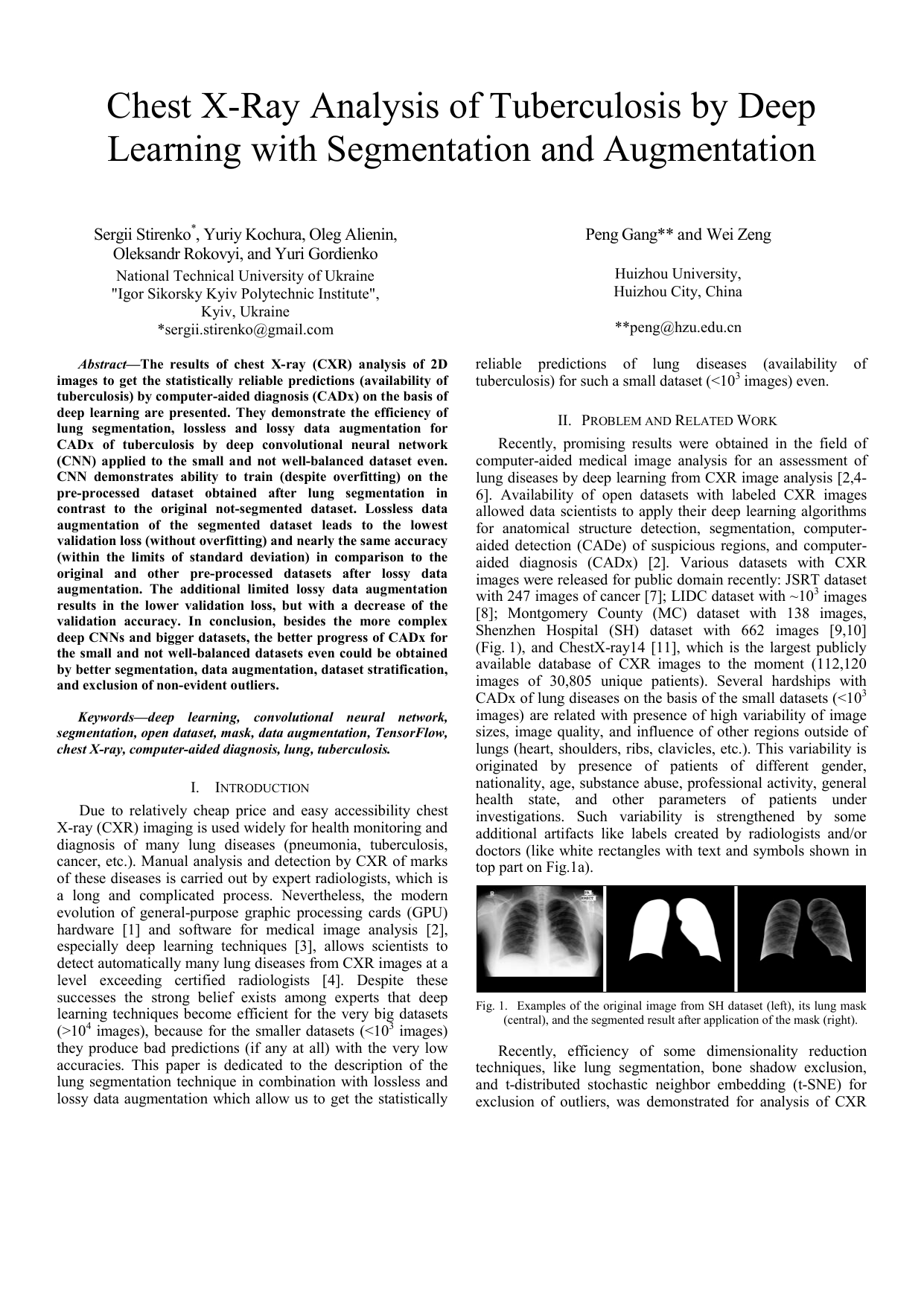
Recently, promising results were obtained in the field of computer-aided medical image analysis for an assessment of lung diseases by deep learning from CXR image analysis [2,[4][5][6]. Availability of open datasets with labeled CXR images allowed data scientists to apply their deep learning algorithms for anatomical structure detection, segmentation, computeraided detection (CADe) of suspicious regions, and computeraided diagnosis (CADx) [2]. Various datasets with CXR images were released for public domain recently: JSRT dataset with 247 images of cancer [7]; LIDC dataset with ~10 3 images [8]; Montgomery County (MC) dataset with 138 images, Shenzhen Hospital (SH) dataset with 662 images [9,10] (Fig. 1), and ChestX-ray14 [11], which is the largest publicly available database of CXR images to the moment (112,120 images of 30,805 unique patients). Several hardships with CADx of lung diseases on the basis of the small datasets (<10 3 images) are related with presence of high variability of image sizes, image quality, and influence of other regions outside of lungs (heart, shoulders, ribs, clavicles, etc.). This variability is originated by presence of patients of different gender, nationality, age, substance abuse, professional activity, general health state, and other parameters of patients under investigations. Such variability is strengthened by some additional artifacts like labels created by radiologists and/or doctors (like white rectangles with text and symbols shown in top part on Fig. 1a). Recently, efficiency of some dimensionality reduction techniques, like lung segmentation, bone shadow exclusion, and t-distributed stochastic neighbor embedding (t-SNE) for exclusion of outliers, was demonstrated for analysis of CXR 2D images to identify marks of lung cancer [5][6]. Despite these attempts, CADx remains a challenge in medical image applications, especially, in the view of high variability of lungs and other regions outside of lungs due to different personal parameters of patients under investigations. The aim of this work is to demonstrate the efficiency of the proposed lung segmentation technique in combination with lossless and lossy data augmentation methods to get the statistically reliable predictions of lung diseases (availability of tuberculosis) even for the relatively small datasets like SH (<10 3 images) [9,10].
The deep CNN [5] was trained on various general-purpose graphic processing cards (GPU) by NVIDIA: Tesla K40 (Kepler microarchitecture), GTX 1080 Ti (Pascal), and the latest and most powerful GPU-card, Tesla V100 (Volta). The official data on performance, bandwidth, number of cores, and the actual training times with the correspondent speedups (for the same dataset and runtime configuration) are shown in Table 1. It worth to note that the CNN model was not optimized here for calculations by tensor processing unit (TPU) in V100 card. That is why its speedup is not so impressive in this work, but the results on the optimized version will be reported elsewhere [12]. Shenzhen Hospital (SH) dataset of CXR images was acquired from Shenzhen No. 3 People’s Hospital in Shenzhen, China. It contains normal and abnormal CXR images with marks of tuberculosis. The exploratory data analysis shown that this small dataset is a not well-balanced dataset with regard to availability/absence of disease, age, gender (Fig. 2). The image widths and heights are not equal and these differences lead to the wide and asymmetric distribution of image areas (Fig. 3). The current and previous attempts to perform training for such small CXR datasets without any pre-processing were performed and failed [5,6]. Fortunately, “external segmentation” of the left and right lung fields in CXR images (exclusion of outside regions whi
This content is AI-processed based on open access ArXiv data.