In vivo wireless medical devices have a critical role in healthcare technologies due to their continuous health monitoring and noninvasive surgery capabilities. In order to fully exploit the potential of such devices, it is necessary to characterize the in vivo wireless communication channel which will help to build reliable and high-performance communication systems. This paper presents preliminary results of experimental characterization for this fascinating communications medium on a human cadaver and compares the results with numerical studies.
2015 IEEE 82nd Vehicular Technology Conference: VTC2015-Fall
6–9 September 2015, Boston, USA
DOI: 10.1109/VTCFall.2015.7390942 1 http://ieeexplore.ieee.org/document/7390942/
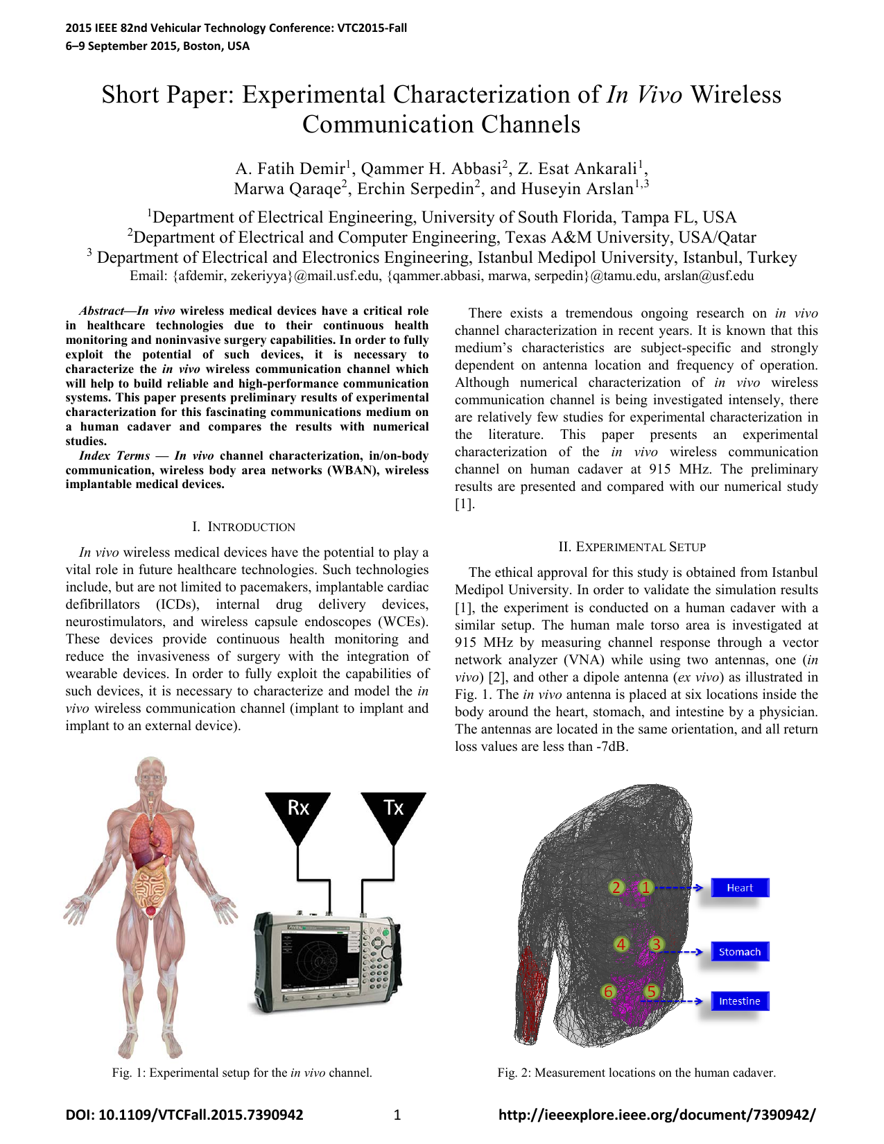
Fig. 2: Measurement locations on the human cadaver.
Fig. 1: Experimental setup for the in vivo channel.
Short Paper: Experimental Characterization of In Vivo Wireless
Communication Channels
A. Fatih Demir1, Qammer H. Abbasi2, Z. Esat Ankarali1,
Marwa Qaraqe2, Erchin Serpedin2, and Huseyin Arslan1,3
1Department of Electrical Engineering, University of South Florida, Tampa FL, USA
2Department of Electrical and Computer Engineering, Texas A&M University, USA/Qatar
3 Department of Electrical and Electronics Engineering, Istanbul Medipol University, Istanbul, Turkey
Email: {afdemir, zekeriyya}@mail.usf.edu, {qammer.abbasi, marwa, serpedin}@tamu.edu, arslan@usf.edu
Abstract—In vivo wireless medical devices have a critical role
in healthcare technologies due to their continuous health
monitoring and noninvasive surgery capabilities. In order to fully
exploit the potential of such devices, it is necessary to
characterize the in vivo wireless communication channel which
will help to build reliable and high-performance communication
systems. This paper presents preliminary results of experimental
characterization for this fascinating communications medium on
a human cadaver and compares the results with numerical
studies.
Index Terms — In vivo channel characterization, in/on-body
communication, wireless body area networks (WBAN), wireless
implantable medical devices.
I. INTRODUCTION
In vivo wireless medical devices have the potential to play a
vital role in future healthcare technologies. Such technologies
include, but are not limited to pacemakers, implantable cardiac
defibrillators
(ICDs),
internal
drug
delivery
devices,
neurostimulators, and wireless capsule endoscopes (WCEs).
These devices provide continuous health monitoring and
reduce the invasiveness of surgery with the integration of
wearable devices. In order to fully exploit the capabilities of
such devices, it is necessary to characterize and model the in
vivo wireless communication channel (implant to implant and
implant to an external device).
There exists a tremendous ongoing research on in vivo
channel characterization in recent years. It is known that this
medium’s characteristics are subject-specific and strongly
dependent on antenna location and frequency of operation.
Although numerical characterization of in vivo wireless
communication channel is being investigated intensely, there
are relatively few studies for experimental characterization in
the
literature.
This
paper
presents
an
experimental
characterization of the in vivo wireless communication
channel on human cadaver at 915 MHz. The preliminary
results are presented and compared with our numerical study
[1].
II. EXPERIMENTAL SETUP
The ethical approval for this study is obtained from Istanbul
Medipol University. In order to validate the simulation results
[1], the experiment is conducted on a human cadaver with a
similar setup. The human male torso area is investigated at
915 MHz by measuring channel response through a vector
network analyzer (VNA) while using two antennas, one (in
vivo) [2], and other a dipole antenna (ex vivo) as illustrated in
Fig. 1. The in vivo antenna is placed at six locations inside the
body around the heart, stomach, and intestine by a physician.
The antennas are located in the same orientation, and all return
loss values are less than -7dB.
2
Table I: Path loss values for selected in vivo locations.
Fig. 4: Path loss versus depth from body surface.
TABLE II: PARAMETERS FOR THE STATISTICAL PATH LOSS MODEL
III. RADIO CHANNEL CHARACTERIZATION
EM wave propagation inside the body is strongly related to
the location of the antenna. Therefore, in vivo wireless channel
characterization is mostly investigated for a specific part of
the human body. Fig. 2 shows the antenna locations inside the
body and Table I summarizes path loss values for these
locations. The signal propagates through different organs and
tissues that the path loss changes significantly for the locations
at similar depth from the body surface.
The channel modeling subgroup (Task Group 15.6), which
worked on developing of the IEEE 802.15.6 standard,
submitted their final report on body area network (BAN)
channel models in November 2010 [3]. In this report, it is
determined that Friis transmission equation can be used for in
vivo scenarios by adding a random variation. In our previous
study [1], the path loss is modeled as a function of depth by
the following equation in dB:
PL (d) = PL0 + m (d / d0) + S (d≥ d0)
where d stands for the depth from body surface in millimeters,
d0 denotes the reference depth (i.e. 10mm), PL0 represents the
inters
This content is AI-processed based on open access ArXiv data.