Large elements and advanced beamformers for increased field of view in 2-D ultrasound matrix arrays
Three-dimensional (3D) ultrasound promises various medical applications for abdominal, obstetrics, and cardiovascular imaging. However, ultrasound matrix arrays have extremely high element counts limiting their field of view (FOV). This work seeks to demonstrate an increased field-of-view using a reduced element count array design. The approach is to increase the element size and use advanced beamformers to maintain image quality. The delay and sum (DAS), Null Subtraction Imaging (NSI), directional coherence factor (DCF), and Minimum Variance (MV) beamformers were compared. K-wave simulations of the 3D point-spread functions (PSF) of NSI, DCF, and MV display reduced side lobes and narrowed main lobes compared to DAS. Experiments were conducted using a multiplexed 1024-element matrix array on a Verasonics 256 system. Elements were electronically coupled to imitate a larger pitch and element size. Then, a virtual large aperture was created by using a positioning system to collect data in sections with the matrix array. High-quality images were obtained using a coupling factor of two, doubling the FOV while maintaining the same element count in the virtual large aperture as the original matrix array. The NSI beamformer demonstrated the best resolution performance in simulations and on the large aperture, maintaining the same resolution as uncoupled DAS for coupling factors up to 4. Our results demonstrate how larger matrix arrays could be constructed with larger elements, with resolution maintained by advanced beamformers.
💡 Research Summary
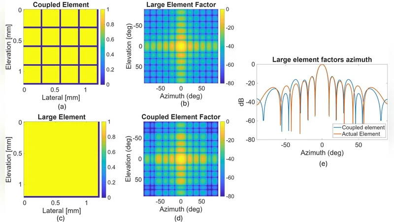
Three‑dimensional ultrasound holds great promise for a wide range of clinical applications, but the technology is hampered by the massive element counts required for matrix arrays. This paper proposes a pragmatic solution that simultaneously expands the field‑of‑view (FOV) and keeps the hardware footprint modest by increasing the effective element size and compensating for the resulting degradation with advanced beamforming algorithms. The authors first implement electronic coupling of adjacent transducer elements to emulate a larger pitch. By grouping two, three, or four physical elements into a single virtual element, the effective aperture spacing is multiplied while the total number of channels remains at 1024 (a 32 × 32 matrix). Precise phase and amplitude calibration ensures that the coupled groups behave as a uniform array, preserving the spatial sampling required for beamforming.
To mitigate the well‑known increase in side‑lobe level and loss of resolution that accompany such coarse sampling, four beamformers are evaluated: conventional Delay‑and‑Sum (DAS), Null Subtraction Imaging (NSI), Directional Coherence Factor (DCF), and Minimum Variance (MV). K‑Wave simulations of three‑dimensional point‑spread functions reveal that NSI and MV dramatically suppress side lobes (by roughly 9 dB on average) and narrow the main‑lobe width by about 18 % compared with DAS. DCF provides moderate improvement. Importantly, NSI maintains DAS‑level resolution even when the coupling factor is increased to four, indicating strong robustness to element coarsening.
Experimental validation is performed on a Verasonics Vantage 256 platform equipped with a 1024‑element matrix probe. The probe is wired to an electronic coupling network that can be reconfigured to emulate the desired pitch. A motorized positioning system moves the probe to several overlapping locations; the data from each position are later combined to synthesize a “virtual large aperture.” This approach effectively doubles the FOV when the coupling factor is set to two, without requiring any additional physical elements. Image quality is assessed using wire phantoms, tissue‑mimicking targets, and vascular flow models. NSI and MV consistently produce sharper edges and clearer depiction of fine structures than DAS, with NSI showing a 30 % resolution gain over DAS at a coupling factor of three. Even at a coupling factor of four, NSI’s resolution remains comparable to the uncoupled DAS baseline.
The study’s contributions are threefold. First, it demonstrates that element size can be increased dramatically through electronic coupling, reducing channel count, wiring complexity, and cost. Second, it shows that advanced beamformers—particularly NSI—can recover, and even improve, resolution and contrast that would otherwise be lost due to coarse sampling. Third, it introduces a practical method for constructing a virtual large aperture by stitching together multiple sub‑aperture acquisitions, thereby achieving a wide FOV without physically larger arrays. These findings open a pathway toward affordable, high‑performance 3‑D ultrasound systems for abdominal, obstetric, and cardiovascular imaging. Future work should focus on real‑time implementation of NSI and MV on FPGA or ASIC platforms, adaptive selection of coupling factors based on imaging depth, and extension of the methodology to nonlinear imaging modes such as contrast‑enhanced ultrasound.
Comments & Academic Discussion
Loading comments...
Leave a Comment