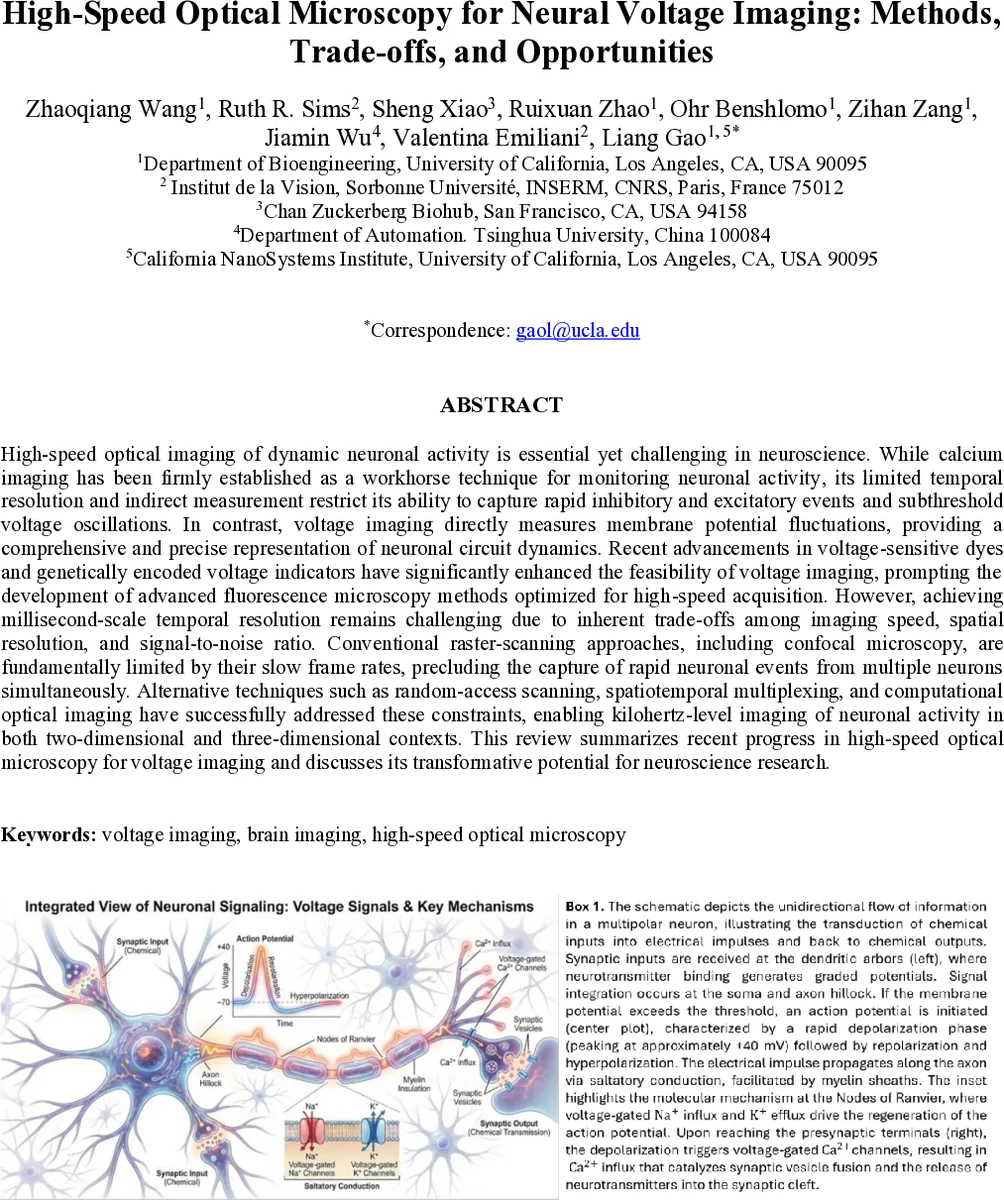
High-speed optical microscopy for neural voltage imaging: Methods, trade-offs, and opportunities
High-speed optical imaging of dynamic neuronal activity is essential yet challenging in neuroscience. While calcium imaging has been firmly established as a workhorse technique for monitoring neuronal activity, its limited temporal resolution and indirect measurement restrict its ability to capture rapid inhibitory and excitatory events and subthreshold voltage oscillations. In contrast, voltage imaging directly measures membrane potential fluctuations, providing a comprehensive and precise representation of neuronal circuit dynamics. Recent advancements in voltage-sensitive dyes and, particularly, genetically encoded voltage indicators have significantly enhanced the feasibility of voltage imaging, prompting the development of advanced fluorescence microscopy methods optimized for high-speed acquisition. However, achieving millisecond-scale temporal resolution remains challenging due to inherent trade-offs among imaging speed, spatial resolution, and signal-to-noise ratio. Conventional raster-scanning approaches, including confocal microscopy, are fundamentally limited by their slow frame rates, precluding the capture of rapid neuronal events from multiple neurons simultaneously. Alternative techniques such as random-access scanning, spatiotemporal multiplexing, and computational optical imaging have successfully addressed these constraints, enabling kilohertz-level imaging of neuronal activity in both two-dimensional and three-dimensional contexts. This review summarizes recent progress in high-speed optical microscopy for voltage imaging and discusses its transformative potential for neuroscience research.
💡 Research Summary
This review provides a comprehensive overview of the state‑of‑the‑art high‑speed optical microscopy techniques that enable direct voltage imaging of neuronal activity. While calcium imaging has become the workhorse for population‑level neural recordings, its intrinsic temporal lag and indirect nature limit its ability to capture rapid inhibitory and excitatory events as well as sub‑threshold membrane dynamics. Voltage imaging, by contrast, reports membrane potential changes on the millisecond timescale, offering a more faithful readout of neuronal computation. However, voltage signals are intrinsically weak because fluorophores are confined to the thin plasma membrane, and the rapid kinetics of action potentials (rise time ≈250 µs) demand frame rates well above 300 Hz, ideally in the kilohertz range. This creates a fundamental trade‑off among imaging speed, spatial resolution, and signal‑to‑noise ratio (SNR).
The authors categorize recent advances into four major methodological families.
-
One‑Photon Planar Imaging – Widefield microscopy provides parallel acquisition of an entire 2‑D field of view, delivering the highest pixel‑throughput. Its main limitation is the lack of optical sectioning, which leads to substantial out‑of‑focus background, especially in densely labeled tissue. Strategies to mitigate this include structured illumination, holographic spot illumination, and targeted excitation of genetically defined neurons. A notable implementation is line‑scan confocal microscopy combined with targeted illumination, which achieved 800 Hz imaging over a 1.1 × 0.325 mm² area, recording from 78 neurons for 20 minutes while improving the signal‑to‑background ratio (SBR) by >50‑fold.
-
Multi‑Depth (Multi‑Z) Confocal Microscopy – By extending illumination axially (low NA) and using a cascade of reflective pinholes, multiple focal planes can be recorded simultaneously. The MuZIC system incorporates an ultrafast polygon scanner (≈55 kRPM) and a secondary linear galvo to scan four planes (128 × 127 pixels each) at 916 Hz. In vivo recordings from mouse motor cortex expressing Voltron2‑ST demonstrated high‑SNR detection of both sub‑threshold and spiking activity at depths of 150–200 µm. Line‑scan variants that replace pinholes with reflective slits further increase throughput, albeit at the cost of some background rejection.
-
Light‑Sheet Fluorescence Microscopy (LSFM) – Light‑sheet approaches illuminate a thin planar section while a camera captures the entire plane in a single exposure, providing optical sectioning with high photon efficiency. Volumetric speed is limited by the axial refocusing mechanism. Remote‑focusing using a lightweight mirror driven by a piezo bender enabled 200.8 Hz volumetric imaging (1.46 µm lateral, 11.7 µm axial resolution) in larval zebrafish brain. The FLIPR (Flipped Image Remote Focusing) design replaces the conventional beamsplitter with a retro‑reflector, doubling light throughput and achieving 500 Hz volumetric imaging over a 150 µm axial range, recording >100 spontaneously active spinal neurons. Parallel camera arrays have pushed LSFM to kilohertz volumetric rates across 250 µm³ volumes, but splitting the emission reduces photon budget per plane and can increase out‑of‑focus background. Single‑objective variants such as SCAPE (Swept‑Confocally‑Aligned Planar Excitation) avoid the orthogonal‑objective constraint and have demonstrated >300 Hz 3‑D imaging of blood flow and calcium transients; although not yet applied to voltage indicators, SCAPE’s geometry is promising for future voltage LSFM.
-
Snapshot Volumetric Imaging – Light‑Field Microscopy (LFM) – LFM records both spatial and angular information via a microlens array, enabling computational refocusing of the entire 3‑D volume from a single camera exposure. Because acquisition speed is limited only by camera readout, LFM can achieve volumetric rates comparable to the frame rate (e.g., 100 vol/s with 1‑photon excitation of sparsely expressed VSFP‑Butterfly 1.2 in mouse brain slices). Digital adaptive optics can be applied post‑acquisition to correct system and sample aberrations without additional hardware, preserving photon efficiency. With advances in high‑quantum‑efficiency cameras and faster GPUs for real‑time reconstruction, kilohertz‑scale light‑field voltage imaging is within reach.
Across all modalities, the central challenge remains the photon budget: increasing excitation power improves SNR but accelerates photobleaching, phototoxicity, and heating. Membrane‑targeted voltage indicators improve signal strength, yet imperfect targeting adds background fluorescence. Moreover, kilohertz imaging generates massive data streams (hundreds of megabytes per second), demanding high‑bandwidth data pipelines, real‑time compression, and substantial storage capacity. Recent progress in deep‑learning‑based denoising, on‑the‑fly compression, and FPGA/GPU‑accelerated processing helps alleviate these bottlenecks.
The review concludes by highlighting emerging opportunities. Continued engineering of brighter, faster, and more specific GEVIs will synergize with optical innovations. Hybrid systems that combine targeted illumination, remote focusing, multi‑camera detection, and computational adaptive optics promise to break the traditional speed‑resolution‑SNR trade‑off, enabling simultaneous kilohertz‑rate, cellular‑resolution, three‑dimensional recordings from thousands of neurons in behaving animals. Such capabilities will open new avenues for probing rapid circuit dynamics, sub‑threshold integration, inhibitory–excitatory balance, and the spatiotemporal propagation of electrical signals that have been inaccessible to conventional electrophysiology or calcium imaging.
Comments & Academic Discussion
Loading comments...
Leave a Comment