In Vivo Measurement of Blood Clot Mechanics from Computational Fluid Dynamics based on Intravital Microscopy Images
Ischemia leading to heart attacks and strokes is the major cause of deaths in the world. Whether an occlusion occurs or not, depends on the ability of a growing thrombus to resist forces exerted on its structure. This manuscript provides the first known in vivo measurement of the stresses that clots can withstand, before yielding to the surrounding blood flow. Namely, Lattice-Boltzmann Method flow simulations are performed based on 3D clot geometries. The latter are estimated from intravital microscopy images of laser-induced injuries in cremaster microvasculature of live mice. In addition to reporting the blood clot yield stresses, we also show that the thrombus ‘core’ does not experience significant deformation, while its ‘shell’ does. This indicates that the latter is more prone to embolization. Hence, drugs should be designed to target the shell selectively, while leaving the core intact (to minimize excessive bleeding). Finally, we laid down a foundation for a nondimensionalization procedure, which unraveled a relationship between clot mechanics and biology. Hence, the proposed framework could ultimately lead to a unified theory of thrombogenesis, capable of explaining all clotting events. Thus, the findings presented herein will be beneficial to the understanding and treatment of heart attacks, strokes and hemophilia.
💡 Research Summary
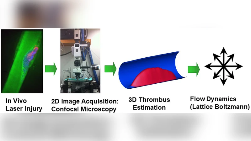
This paper presents the first in‑vivo quantification of the mechanical stresses that a growing thrombus can withstand before yielding to the surrounding blood flow. Using a well‑established mouse cremaster model, the authors induced localized vascular injury with a focused laser and captured the ensuing thrombus formation in real time with intravital microscopy. Two fluorescence channels were recorded simultaneously: one labeling fibrin (core) and the other labeling activated platelets (shell). After denoising and edge detection, the sequential 2‑D image stacks were volumetrically reconstructed to obtain a high‑resolution three‑dimensional geometry of each clot. The geometry was then discretized onto a lattice for computational fluid dynamics (CFD) simulations based on the Lattice‑Boltzmann Method (LBM), which is particularly suited for complex moving boundaries and non‑Newtonian blood rheology.
The CFD model incorporated experimentally measured inlet velocity profiles, vessel diameter, and a Herschel‑Bulkley viscosity model for whole blood. By solving the LBM equations, the authors obtained spatially resolved distributions of shear stress (τ) and normal stress (σ) on the clot surface throughout the cardiac cycle. The stress field was mapped onto the previously defined core‑shell regions. The core, composed of a dense fibrin network and tightly packed platelets, experienced shear stresses below ~0.5 Pa and displayed negligible deformation. In contrast, the peripheral shell, directly exposed to the lumen, was subjected to higher shear stresses; once τ exceeded an empirically determined threshold of approximately 1.2 Pa, the shell exhibited rapid strain accumulation and occasional detachment of fragments, indicating a high propensity for embolization.
To generalize these findings across different vascular scales and flow conditions, the authors introduced a nondimensionalization scheme. They defined dimensionless groups analogous to Reynolds (Re), Weber (We), and a shear‑stress number (τ*), and performed regression analysis linking τ* to biological markers such as plasmin activity and fibrin density. Remarkably, data from vessels ranging from 10 µm to 100 µm in diameter collapsed onto a single master curve, suggesting that the identified stress threshold is robust across physiologically relevant geometries.
The biological implication of the core‑shell mechanical dichotomy is profound. The core’s mechanical integrity is essential for hemostasis, whereas the shell’s susceptibility to flow‑induced deformation makes it the primary source of downstream emboli. Consequently, the authors argue that antithrombotic therapies should be refined to selectively target the shell while preserving the core, thereby reducing the risk of excessive bleeding. Potential strategies include shell‑specific drug delivery vehicles (e.g., nanoparticles functionalized with antibodies against activated platelet markers) or localized release of fibrinolytics confined to the shell region.
Beyond the immediate clinical relevance, the study establishes a methodological pipeline that integrates high‑resolution intravital imaging, 3‑D geometric reconstruction, LBM‑based CFD, and nondimensional analysis. This pipeline can be adapted to human imaging data (e.g., OCT or high‑resolution CT) and extended to evaluate the mechanical efficacy of novel antithrombotic agents before in‑vivo testing. By providing a quantitative link between clot biology and mechanics, the work moves the field toward a unified theory of thrombogenesis capable of predicting when a clot will remain stable, when it will grow, and when it will break apart to cause stroke or myocardial infarction.
In summary, the paper delivers (1) the first direct measurement of in‑vivo clot yield stress, (2) a clear mechanical distinction between core and shell regions, (3) a nondimensional framework that unifies clot mechanics across scales, and (4) actionable insights for designing shell‑targeted antithrombotic therapies. These contributions have the potential to reshape both basic research on hemostasis and the clinical management of thrombotic diseases.
Comments & Academic Discussion
Loading comments...
Leave a Comment