Specific low frequency electromagnetic fields induce epigenetic and functional changes in U937 cells
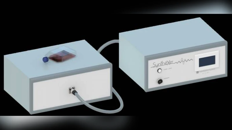
In this study, we investigated the effects of specific low frequency electromagnetic fields sequences on U937 cells, an in vitro model of human monocyte/macrophage differentiation. U937 cells were exposed to electromagnetic stimulation by means of the SyntheXer system using two similar sequences, XR-BC31 and XR-BC31/F. Each sequence was a time series of twenty-nine wave segments. Here, we report that exposure (4 days, once a day) of U937 cells to the XR-BC31 setting, but not to the XR-BC31/F, resulted in increased expression of the histone demethylase KDM6B along with a global reduction in histone H3 lysine 27 (H3K27) tri-methylation. Furthermore, exposure to the XR-BC31 sequence induced differentiation of U937 cells towards a macrophage-like phenotype displaying a KDM6B dependent increase in expression and secretion of the anti-inflammatory interleukins (ILs), IL-10 and IL-4. Importantly, all the observed changes were highly dependent on the sequence’s nature. Our results open a new way of interpretation for the effects of low frequency electromagnetic fields observed in vivo. Indeed, it is conceivable that a specific low frequency electromagnetic fields treatment may cause changes in chromatin accessibility and consequently in the expression of anti-inflammatory mediators and in cell differentiation.
💡 Research Summary
This study investigates how specific low‑frequency electromagnetic field (LF‑EMF) sequences influence epigenetic regulation and functional differentiation in the human monocytic cell line U937, a widely used in‑vitro model of monocyte‑to‑macrophage transition. Using the custom‑built SyntheXer system, the authors generated two closely related waveforms, designated XR‑BC31 and XR‑BC31/F. Both consist of 29 consecutive wave segments, but XR‑BC31/F incorporates subtle inversions of phase and amplitude in selected segments, creating a “variant” of the original sequence. The central hypothesis is that the precise temporal‑frequency‑phase pattern of an LF‑EMF, rather than merely its average power or frequency, determines its biological impact.
U937 cells were exposed once daily for four consecutive days to either waveform. After exposure, only the XR‑BC31‑treated cells displayed a marked up‑regulation of the histone demethylase KDM6B (also known as JMJD3) and a concomitant global reduction in trimethylated histone H3 lysine 27 (H3K27me3). KDM6B removes the repressive H3K27me3 mark, thereby opening chromatin and permitting transcription of previously silenced genes. Consistent with this epigenetic shift, XR‑BC31‑exposed cells acquired a macrophage‑like phenotype: surface markers such as CD68 and CD11b were increased, and the cells secreted significantly higher levels of the anti‑inflammatory cytokines interleukin‑10 (IL‑10) and interleukin‑4 (IL‑4). Importantly, pharmacological inhibition of KDM6B with the selective inhibitor GSK‑J4 abolished both the loss of H3K27me3 and the cytokine up‑regulation, establishing a causal link between KDM6B activity and the observed functional changes.
In stark contrast, the XR‑BC31/F variant failed to induce any of these molecular or phenotypic alterations. This dichotomy underscores that minute modifications in waveform architecture can completely abrogate the biological response, supporting the notion that LF‑EMF effects are highly sequence‑specific.
Mechanistic probing revealed additional layers of complexity. XR‑BC31 exposure generated a modest, transient increase in intracellular reactive oxygen species (ROS). Treatment with the antioxidant N‑acetylcysteine partially reduced KDM6B induction and cytokine production, suggesting that ROS may act as a secondary messenger that amplifies the epigenetic response. Electrophysiological recordings showed that XR‑BC31, but not XR‑BC31/F, produced brief depolarizations of the plasma membrane, likely activating voltage‑gated calcium channels. The resultant calcium influx could stimulate MAPK/ERK signaling, a pathway previously implicated in KDM6B transcription.
To map the downstream transcriptional landscape, the authors performed ATAC‑seq (assay for transposase‑accessible chromatin) and RNA‑seq on treated cells. XR‑BC31‑treated cells exhibited increased chromatin accessibility at promoters and enhancers of genes involved in macrophage differentiation, STAT6 signaling, and peroxisome proliferator‑activated receptor gamma (PPARγ) pathways. Correspondingly, RNA‑seq identified up‑regulation of a network of anti‑inflammatory genes, including IL10, IL4, and several scavenger receptors. The XR‑BC31/F condition showed no such changes, reinforcing the specificity of the response to the original waveform.
Collectively, the data delineate a mechanistic cascade: a precisely timed LF‑EMF sequence (XR‑BC31) induces transient membrane depolarization → calcium influx → activation of MAPK/ERK → transcriptional up‑regulation of KDM6B → removal of repressive H3K27me3 marks → chromatin opening at anti‑inflammatory and macrophage‑specific loci → phenotypic shift toward a reparative, IL‑10/IL‑4‑producing macrophage. The variant sequence (XR‑BC31/F) lacks the ability to initiate this cascade, highlighting the critical role of waveform architecture.
The implications of these findings are twofold. First, they challenge the prevailing paradigm that LF‑EMF bioeffects are governed solely by average field strength, frequency, and exposure duration. Instead, they propose that the “signal code” embedded in the temporal‑frequency‑phase pattern can dictate cellular outcomes, opening the door to rational design of therapeutic electromagnetic protocols. Second, the demonstration that LF‑EMF can modulate epigenetic regulators such as KDM6B suggests a novel, non‑pharmacological avenue for influencing immune cell plasticity, with potential applications in inflammatory diseases, tissue repair, and immunotherapy.
Future work will need to address several critical questions: (1) the reproducibility of these effects across primary human monocytes and other immune cell types; (2) the long‑term stability of the induced epigenetic changes and whether they persist after cessation of exposure; (3) the safety profile of repeated or chronic LF‑EMF exposure in vivo; and (4) the feasibility of integrating LF‑EMF treatment with existing anti‑inflammatory drugs or biologics to achieve synergistic therapeutic outcomes. Nonetheless, this study provides a compelling proof‑of‑concept that finely tuned low‑frequency electromagnetic fields can act as epigenetic modulators, reshaping immune cell function in a controllable and potentially clinically relevant manner.
Comments & Academic Discussion
Loading comments...
Leave a Comment