Molecular Dynamics Simulation of Vascular Network Formation
Endothelial cells are responsible for the formation of the capillary blood vessel network. We describe a system of endothelial cells by means of two-dimensional molecular dynamics simulations of point-like particles. Cells’ motion is governed by the gradient of the concentration of a chemical substance that they produce (chemotaxis). The typical time of degradation of the chemical substance introduces a characteristic length in the system. We show that point-like model cells form network resembling structures tuned by this characteristic length, before collapsing altogether. Successively, we improve the non-realistic point-like model cells by introducing an isotropic strong repulsive force between them and a velocity dependent force mimicking the observed peculiarity of endothelial cells to preserve the direction of their motion (persistence). This more realistic model does not show a clear network formation. We ascribe this partial fault in reproducing the experiments to the static geometry of our model cells that, in reality, change their shapes by elongating toward neighboring cells.
💡 Research Summary
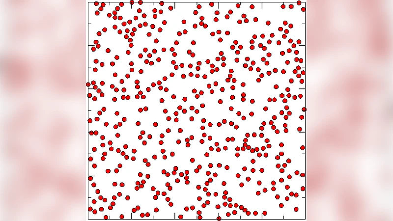
The paper investigates the physical mechanisms underlying capillary network formation by endothelial cells using two‑dimensional molecular dynamics (MD) simulations. In the first, simplest model, each cell is represented as a point‑like particle that moves up the gradient of a chemoattractant it secretes. The chemoattractant diffuses with coefficient D and degrades after a characteristic time τ, which introduces a length scale λ = √(Dτ). Simulations show that when inter‑cellular distances are smaller than λ, cells aggregate into transient lattice‑like or filamentous patterns, but the structures eventually collapse into a single cluster or disperse, reflecting the inability of a point‑particle description to capture volume exclusion and shape change.
To address these shortcomings, the authors augment the model with two biologically motivated forces. First, an isotropic short‑range repulsion prevents particles from overlapping and enforces a minimum separation r₀, mimicking physical cell volume. Second, a velocity‑dependent “persistence” force penalizes rapid changes in direction, reflecting the experimentally observed tendency of endothelial cells to maintain their migration direction over time. The persistence term is implemented as a cosine‑type coupling between the current velocity vector and the previous heading.
When both forces are included, the system initially forms small clusters that respect the imposed exclusion distance. However, as simulation time progresses, clusters either merge or dissolve, and no stable, branched network emerges. The authors attribute this failure to the static, spherical representation of cells. In reality, endothelial cells elongate, extend filopodia, and remodel their shape in response to neighboring cells, thereby dynamically adjusting both mechanical contacts and chemotactic cues. These shape changes are essential for the long‑range connectivity and robustness of vascular networks observed in vitro and in vivo.
The study therefore draws several key conclusions. (1) Chemotaxis alone can generate early‑stage patterning, but the characteristic degradation time of the chemoattractant sets a length scale that limits pattern persistence. (2) Adding volume exclusion and directional persistence improves realism but does not suffice to reproduce stable networks when cells are constrained to remain spherical. (3) The omission of dynamic cell morphology—particularly elongation toward neighbors and the associated mechanotransduction pathways—is the primary reason for the discrepancy with experimental observations.
Future work should incorporate deformable cell models, explicit cell‑cell adhesion (e.g., VE‑cadherin mediated bonds), and intracellular signaling networks (such as Rho‑GTPase–actin dynamics) that regulate shape and traction forces. By coupling these mechanical and biochemical degrees of freedom, simulations could capture the self‑organizing behavior of endothelial cells that leads to persistent, functional vascular plexus formation.
Comments & Academic Discussion
Loading comments...
Leave a Comment