Cellular Fourier analysis for geometrically disordered materials
Many media are divided into elementary units with irregular shape and size, as exemplified by domains in magnetic materials, bubbles in foams, or cells in biological tissues. Such media are essentially characterized by geometrical disorder of their elementary units, which we term cells. Cells set a reference scale at which are often assessed parameters and fields reflecting material properties and state. Here, we consider the spectral analysis of spatially varying fields. Such analysis is difficult in geometrically disordered media, because space discretization based on standard coordinate systems is not commensurate with the natural discretization into geometrically disordered cells. Indeed, we found that two classical spectral methods, the Fast Fourier Transform and the Graph Fourier transform, fail to reproduce all expected properties of spectra of plane waves and of white noise. We therefore built a method, which we call Cellular Fourier Transform (CFT), to analyze cell-scale fields, which comprise both discrete fields defined only at cell level and continuous fields smoothed out from their sub-cell variations. Our approach is based on the construction of a discrete operator suited to the disordered geometry and on the computation of its eigenvectors, which, respectively, play the same role as the Laplace operator and sine waves in Euclidean coordinate systems. We show that CFT has the expected behavior for sinusoidal fields and for random fields with long-range correlations. Our approach for spectral analysis is suited to any geometrically disordered material, such as a biological tissue with complex geometry, opening the path to systematic multiscale analyses of material behavior.
💡 Research Summary
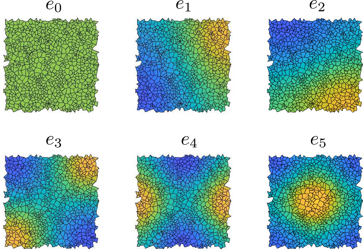
The paper addresses a fundamental challenge in the spectral analysis of spatial fields defined on media whose elementary units—referred to as “cells”—are highly irregular in shape, size, and connectivity. Typical examples include magnetic domains, bubbles in foams, and biological cells. Conventional spectral tools such as the Fast Fourier Transform (FFT) and the Graph Fourier Transform (GFT) rely on regular Cartesian grids or simple graph representations, respectively. When applied to geometrically disordered cellular structures, both methods fail to reproduce the expected spectral properties of elementary signals such as plane waves and white noise. FFT suffers from windowing and low‑pass filtering effects caused by the mismatch between the regular grid and the irregular cell layout, leading to a systematic decay of amplitude with increasing wave number. GFT, which builds a Laplacian on a graph whose vertices are cells and edges encode neighbor relations, loses essential geometric information (cell area, boundary length, shape) and therefore cannot establish a clear correspondence between eigenvalues and physical wave numbers; its spectra for white noise also display a spurious decreasing trend.
To overcome these limitations, the authors develop the Cellular Fourier Transform (CFT), a framework that respects the full geometry of the cellular tessellation. The construction proceeds in several steps. First, a continuous field (f(\mathbf{x})) defined over the domain (\Omega) is “cellularized” by averaging it over each cell (\omega_i), yielding a piecewise‑constant field (f_C). This discretization uses an orthonormal basis (\psi_i(\mathbf{x}) = 1/\sqrt{\mu_i}) inside cell (\omega_i) (zero elsewhere), where (\mu_i) is the cell’s measure (area in 2‑D). The vector of coefficients (f_i) thus represents the field in an (N)-dimensional representation space (with (N) cells).
Next, the authors introduce a coarse, translation‑invariant integral operator that mimics the continuous Laplacian: \
Comments & Academic Discussion
Loading comments...
Leave a Comment