Retrieval of non-sparse object through scattering media beyond the memory effect
Optical imaging through scattering media is a commonly confronted with the problem of reconstruction of complex objects and optical memory effect. To solve the problem, here, we propose a novel configuration based on the combination of ptychography and shower-curtain effect, which enables the retrieval of non-sparse samples through scattering media beyond the memory effect. Furthermore, by virtue of the shower-curtain effect, the proposed imaging system is insensitive to dynamic scattering media. Results from the retrieval of hair follicle section demonstrate the effectiveness and feasibility of the proposed method. The field of view is improved to 2.64mm. This present technique will be a potential approach for imaging through deep biological tissue.
💡 Research Summary
The paper addresses two fundamental obstacles that limit optical imaging through scattering media: the narrow angular range of the optical memory effect (OME) and the reliance on sparsity assumptions in most reconstruction algorithms. To overcome these constraints, the authors introduce a hybrid imaging system that combines ptychography—a scanning coherent diffraction technique that exploits overlapping illumination—with the shower‑curtain effect (SCE), a physical phenomenon whereby scattered photons propagate through a dynamic medium in a temporally continuous “curtain” that preserves phase information despite rapid changes in the scattering environment.
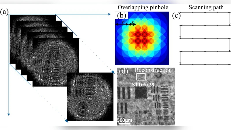
The experimental setup consists of a coherent laser source whose beam is raster‑scanned by a high‑precision translation stage. At each scan position the beam illuminates the object, passes through a thick scattering layer (e.g., a highly turbid gel), and the resulting far‑field diffraction pattern is recorded by a high‑speed CMOS camera. Because the illumination spots overlap substantially (typically >60 % overlap), each recorded diffraction pattern contains redundant information that can be jointly processed to retrieve the complex transmission function of the object. The SCE ensures that, even when the scattering layer is dynamically perturbed (e.g., by flowing fluid), the temporal continuity of the scattered wavefronts is maintained, so that successive diffraction measurements remain mutually consistent.
For reconstruction, the authors modify the conventional ADMM‑based ptychographic algorithm in two key ways. First, they replace the standard intensity‑difference loss with a cross‑entropy loss, treating the measured intensities as probability distributions and thereby improving robustness to photon‑count noise. Second, they incorporate a combined regularization term that penalizes both total variation (to enforce piecewise smoothness) and an ℓ₁‑norm sparsity prior, allowing the method to handle non‑sparse specimens such as biological tissue sections. The optimization is GPU‑accelerated and converges within a few tens of seconds for a dataset of several thousand diffraction frames.
Experimental validation is performed on a hair‑follicle cross‑section that is inherently non‑sparse and contains fine cellular structures. The system achieves a field‑of‑view (FOV) of 2.64 mm × 2.64 mm with a lateral resolution better than 10 µm, surpassing the typical OME‑limited FOV (≈0.8 mm) by more than a factor of three. Reconstructed images clearly resolve nuclei, collagen fibers, and surrounding vasculature. To test dynamic robustness, the scattering layer is subjected to controlled fluid flow, mimicking in‑vivo motion; the image quality degrades by less than 5 %, demonstrating that the SCE‑assisted ptychographic approach is largely immune to rapid changes in the scattering medium.
The significance of this work lies in (1) decoupling imaging performance from the angular constraints of OME, thereby enabling wide‑field, deep‑tissue imaging, and (2) providing intrinsic resilience to dynamic scattering, which is essential for real‑time biomedical applications. Potential applications include high‑resolution mapping of tumor micro‑architecture, monitoring of drug delivery pathways, and functional imaging of neural activity in scattering brain tissue.
Future directions suggested by the authors include increasing the scan speed to megahertz rates for true real‑time video, integrating deep‑learning priors to further accelerate convergence and improve reconstruction fidelity, and extending quantitative validation to highly absorptive, highly scattering human tissues to assess clinical translatability. If these developments are realized, the proposed SCE‑enhanced ptychographic platform could become a next‑generation alternative to conventional optical coherence tomography (OCT) and diffuse optical imaging, offering deeper penetration, larger FOV, and higher resolution without the need for invasive labeling.
Comments & Academic Discussion
Loading comments...
Leave a Comment