GAN-based Multiple Adjacent Brain MRI Slice Reconstruction for Unsupervised Alzheimers Disease Diagnosis
Unsupervised learning can discover various unseen diseases, relying on large-scale unannotated medical images of healthy subjects. Towards this, unsupervised methods reconstruct a single medical image to detect outliers either in the learned feature …
Authors: Changhee Han, Leonardo Rundo, Kohei Murao
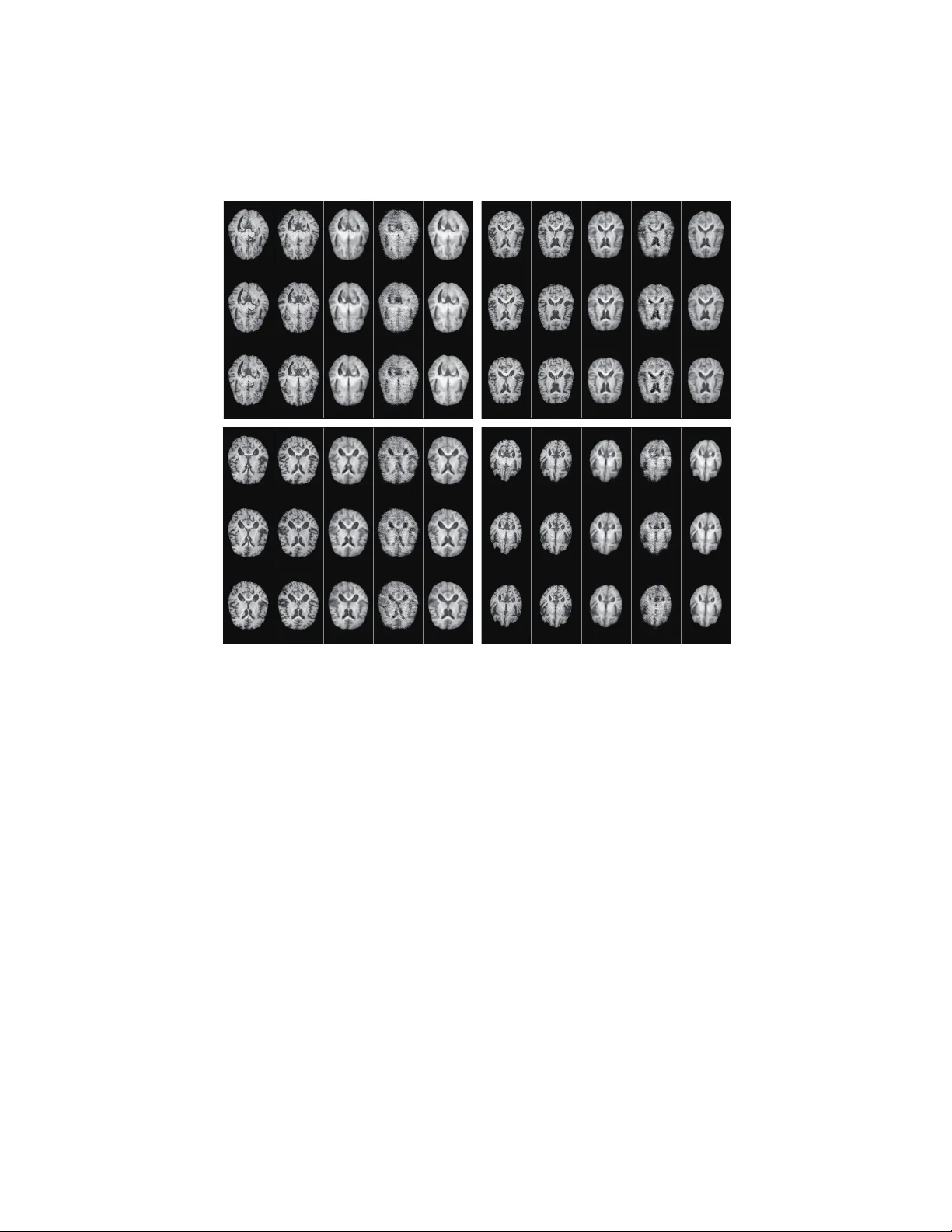
GAN-Based Multiple Adjacen t Brain MRI Slice Reconstruction for Unsup ervised Alzheimer’s Disease Diagnosis Changhee Han 1 , 2 , 3 ( ), Leonardo Rundo 4 , 5 , Kohei Murao 1 , Zolt´ an ´ Ad´ am Milacski 6 , Kazuki Umemoto 7 , Evis Sala 4 , 5 , Hideki Nak a yama 3 , 8 , and Shin’ic hi Satoh 1 1 Researc h Cen ter for Medical Big Data, National Institute of Informatics, T oky o, Japan han@nlab.ci.i.u-tokyo.ac.jp , 2 LPixel Inc., T oky o, Japan 3 Graduate School of Information Science and T ec hnology , The Universit y of T oky o, T oky o, Japan 4 Departmen t of Radiology , Universit y of Cambridge, Cam bridge, United Kingdom 5 Cancer Research UK Cambridge Cen tre, Cam bridge, United Kingdom 6 Departmen t of Artificial Intelligence, EL TE E¨ otv¨ os Lor´ and Univ ersity , Budap est, Hungary 7 Departmen t of Rehabilitation Medicine, Jun tendo Univ ersity Sc ho ol of Medicine, T okyo, Japan 8 In ternational Researc h Cen ter for Neuroin telligence (WPI-IR CN), The Univ ersity of T okyo Institutes for Adv anced Study , The Universit y of T okyo, T oky o, Japan Abstract. Unsup ervised learning can disco ver v arious unseen diseases, relying on large-scale unannotated medical images of health y sub jects. T ow ards this, unsupervised metho ds reconstruct a single medical image to detect outliers either in the learned feature space or from high recon- struction loss. Ho w ever, without considering con tinuit y betw een multiple adjacen t slices, they cannot directly discriminate diseases comp osed of the accumulation of subtle anatomical anomalies, suc h as Alzheimer’s Disease (AD). Moreo ver, no study has sho wn how unsupervised anomaly detection is asso ciated with disease stages. Therefore, w e propose a t wo- step metho d using Generativ e Adversarial Netw ork-based multiple adja- cen t brain MRI slice reconstruction to detect AD at v arious stages: ( R e- c onstruction ) W asserstein loss with Gradien t P enalty + ` 1 loss—trained on 3 healthy slices to reconstruct the next 3 ones—reconstructs unseen health y/AD cases; ( Diagnosis ) Av erage/Maximum loss (e.g., ` 2 loss) per scan discriminates them, comparing the reconstructed/ground truth im- ages. The results show that we can reliably detect AD at a very early stage with Area Under the Curve (A UC) 0 . 780 while also detecting AD at a late stage muc h more accurately with AUC 0 . 917; since our metho d is fully unsup ervised, it should also discov er and alert any anomalies including rare disease. Keyw ords: Generative adversarial net w orks · Alzheimer’s disease diag- nosis · Unsup ervised anomaly detection · Brain MRI reconstruction. 2 C. Han et al. 1 In tro duction Deep Learning can ac hieve accurate computer-assisted diagnosis when large-scale annotated training samples are a v ailable. In medical imaging, unfortunately , preparing suc h massive annotated datasets is often unfeasible; to tac kle this imp ortan t problem, researc hers hav e prop osed v arious data augmentation tec h- niques, including Generativ e Adversarial Netw ork (GAN)-based ones [1,2,3,4,5]. Ho wev er, even exploiting these techniques, sup ervised learning still requires man y images with pathological features, even for rare disease, to make a reliable diagnosis; nevertheless, it can only detect already-learned sp ecific pathologies. In this regard, as physicians notice previously unseen anomaly examples using prior information on healthy b o dy structure, unsup ervised anomaly detection metho ds leveraging only large-scale healthy images can disco ver and alert un- seen disease when their generalization fails. T o wards this, researc hers reconstructed a single medical image via GANs [6], AutoEnco ders (AEs) [7], or com bining them, since GANs can generate realistic images and AEs, esp ecially V ariational AEs, can directly map data onto its latent represen tation [8]; then, unseen images were scored by comparing them with re- constructed ones to discriminate a pathological image distribution (i.e., outliers either in the learned feature space or from high reconstruction loss). How ev er, those single image reconstruction metho ds mainly target diseases easy-to-detect from a single image even for non-exp ert h uman observers, suc h as glioblastoma on Magnetic Resonance (MR) images [8] and lung cancer on Computed T o- mograph y images [7]. Without considering contin uit y b et ween m ultiple adjacent images, they cannot directly discriminate diseases comp osed of the accum ulation of subtle anatomical anomalies, such as Alzheimer’s Disease (AD). Moreov er, no study has sho wn so far ho w unsup ervised anomaly detection is asso ciated with disease stages. W e thus propose a tw o-step metho d using GAN-based multiple adjacen t brain MRI slice reconstruction to detect AD at v arious stages (Fig. 1): ( R e c onstruction ) W asserstein loss with Gradient Penalt y (W GAN-GP) [9,10] + ` 1 loss—trained on 3 healthy brain axial MRI slices to reconstruct the next 3 ones—reconstructs unseen health y/AD cases; ( Diagnosis ) Av erage/Maximum loss (e.g., ` 2 loss) p er scan discriminates them, comparing the reconstructed and ground truth images. Contributions. Our main con tributions are as follows: – MRI Slice Reconstruction: This first m ultiple MRI slice reconstruction approac h can predict the next 3 brain MRI slices from the previous 3 ones only for unseen images similar to training data b y combining WGAN-GP and ` 1 loss. – Unsup ervised Anomaly Detection: This first unsup ervised anomaly de- tection across different disease stages reveals that, like physicians’ wa y of diagnosis, massive health y data can reliably aid early diagnosis, such as of MCI, while also detecting late-stage disease muc h more accurately b y dis- criminating with ` 2 loss. GAN-Based Unsup ervised Alzheimer’s Disease Diagnosis 3 T rain Infer T r ain GAN to reconstruct next 3 healthy MRI slices from previous 3 ones Based on reconstr uction, c lassify MRI scans into healthy / Alzhheimer’s disease Unseen 3 slices Next 3 slices Compare average/maximum loss per scan Reconstructed 3 slices T r ain Inf er T r ain GAN to r econstr uct ne xt 3 healthy MRI slices fr om pr e vious 3 ones Based on r econstr uction, c lassify MRI scans into healthy / Alzh heimer’s disease Unseen 3 slices Ne xt 3 slices Compar e a v er a ge/maximum loss per scan R econstr ucted 3 slices Fig. 1. Unsup ervised AD diagnosis framework: w e train W GAN-GP + ` 1 loss on 3 health y brain axial MRI slices to reconstruct the next 3 ones, and test it on b oth unseen healthy and AD cases to classify them based on a verage/maxim um loss (e.g., ` 2 loss) p er scan. – Alzheimer’s Disease Diagnosis: This first unsup ervised AD diagnosis study can reliably detect AD and also an y other diseases. The remainder of the manuscript is organized as follo ws: Sect. 2 outlines the state-of-the-art of automated AD diagnosis; Sect. 3 describ es the analyzed MRI dataset, as well as the proposed GAN-based unsup ervised AD diagnosis frame- w ork; exp erimen tal results are shown and discussed in Sect. 4; finally , Sect. 5 pro vides conclusive remarks and future w ork. 2 Automated Alzheimer’s Disease Diagnosis Despite the clinical, so cial, and economic significance of early AD diagnosis— primarily asso ciated with MCI detection—it generally relies on sub jective as- sessmen t b y physicians (e.g., neurologists, geriatricians, and psychiatrists); to tac kle this open challenge, researc hers hav e used classic sup ervised Machine Learning based on hand-crafted features [11,12]. More recently , Deep Learn- ing has attracted great attentions owing to its more abstract and descriptiv e em b edding based on multiple non-linear transformations: Liu et al. used a semi- sup ervised CNN to significan tly reduce the need for lab eled training data[13]; for clinical decision-making, Suk et al. integrated multiple sparse regression mo dels (namely , Deep Ensemble Sparse Regression Netw ork) [14]; Spaso v et al. devised a parameter-efficien t CNN for 3D separable conv olutions, combining dual learning and a specific la yer to predict the con v ersion from MCI to AD within 3 y ears [15]; 4 C. Han et al. instead of exploiting the CNNs, Parisot used a semi-sup ervised Graph Con volu- tional Net work trained on a sub-set of lab eled no des with diagnostic outcomes to represen t sparse clinical data [16]. Ho wev er, to the b est of our knowledge, no existing work has conducted fully unsup ervised anomaly detection for AD diagnosis since capturing subtle anatom- ical differences b et w een MCI and AD is challenging. 3 Materials and Metho ds 3.1 O ASIS-3 Da taset W e use a longitudinal dataset of 176 × 240/176 × 256 T1-weigh ted (T1w) 3T brain axial MRI slices containing b oth normal aging sub jects/AD patien ts ex- tracted from the Op en Access Series of Imaging Studies-3 (O ASIS-3) [17]. The 176 × 240 slices are zero-padded to reach 176 × 256 pixels. Relying on Clinical Demen tia Rating (CDR) [18], common clinical scale for the staging of dementia, the sub jects are comprised of: – Unc hanged CDR = 0: Cognitively health y p opulation; – CDR = 0 . 5: V ery mild demen tia ( ∼ MCI); – CDR = 1: Mild demen tia; – CDR = 2: Mo derate dementia. Since our dataset is longitudinal and the same sub ject’s CDRs may v ary (e.g., CDR = 0 to CDR = 0 . 5), we only use scans with unchanged CDR = 0 to assure certainly healthy scans. As CDRs and MRI scans are not alw ays simultaneously acquired, we lab el MRI scans with CDRs at the closest date. W e only select brain MRI slices including hippo campus/amygdala/v en tricles among whole 256 axial slices p er scan to a void ov er-fitting from AD-irrelev an t information; the atroph y of the hipp ocampus/amygdala/cerebral cortex, and enlarged ven tricles are strongly asso ciated with AD, and thus they mainly affect the AD classifi- cation p erformance of Machine Learning [19]. Moreo ver, w e discard lo w-quality MRI slices. The remaining dataset is divided as follows: – T raining set: Unc hanged CDR = 0 (408 sub jects/1 , 133 scans/57 , 834 slices); – V alidation set: Unc hanged CDR = 0 (55 sub jects/155 scans/8 , 080 slices), CDR = 0 . 5 (53 sub jects/85 scans/4 , 607 slices), CDR = 1 (29 sub jects/45 scans/2 , 518 slices), CDR = 2 (2 sub jects/4 scans/160 slices); – T est set: Unc hanged CDR = 0 (113 sub jects/318 scans/16 , 198 slices), CDR = 0 . 5 (99 sub jects/168 scans/9 , 206 slices), CDR = 1 (61 sub jects/90 scans/5 , 014 slices), CDR = 2 (4 sub jects/6 scans/340 slices). The same s ub ject’s scans are included in the same dataset. The datasets are strongly biased tow ards healthy scans similarly to MRI insp ection in the clin- ical routine. During training for reconstruction, we only use the training set con taining healthy slices to conduct unsupervised learning. GAN-Based Unsup ervised Alzheimer’s Disease Diagnosis 5 3.2 GAN-based Multiple Adjacent Brain MRI Slice Reconstruction T o mo del strong consistency in healthy brain anatomy (Fig. 1), in each scan, we reconstruct the next 3 MRI slices from the previous 3 ones using an image-to- image GAN (e.g., if a scan includes 40 slices s i for i = 1 , . . . , 40, we reconstruct all p ossible 35 setups: ( s i ) i ∈{ 1 , 2 , 3 } 7→ ( s i ) i ∈{ 4 , 5 , 6 } ; ( s i ) i ∈{ 2 , 3 , 4 } 7→ ( s i ) i ∈{ 5 , 6 , 7 } ; . . . ; ( s i ) i ∈{ 35 , 36 , 37 } 7→ ( s i ) i ∈{ 38 , 39 , 40 } ). W e concatenate adjacen t 3 grayscale slices in to 3 c hannels, suc h as in RGB images. The GAN uses a U-Net-lik e [20,21] generator with 4 con volutional lay ers in enco ders and 4 deconv olutional lay ers in decoders resp ectiv ely with skip connections as well as a discriminator with 3 deco ders. W e apply batc h normalization to b oth con volution with Leaky Rectified Linear Unit (ReLU) and deconv olution with ReLU. T o confirm ho w reconstructed im- ages’ realism and anatomical contin uity affect anomaly detection, w e compare the GAN mo dels with different loss functions: ( i ) Dice loss (i.e., a plain U-Net without the discriminator); ( ii ) W GAN-GP loss; ( iii ) WGAN-GP loss + 100 ` 1 loss. Among 8 losses comparing ground truth/reconstructon, a verage ` 2 loss p er scan alwa ys outp erforms the other losses during v alidation for U-Net and W GAN-GP without/with ` 1 loss, and th us we use this loss for testing. Implementation Details Considering its computational sp eed, U-Net training lasts for 600 , 000 steps with a batc h size of 64 and b oth GAN trainings last for 300 , 000 steps with a batch size of 32. W e use 2 . 0 × 10 − 4 learning rate for the Adam optimizer [22]. The framework is implemented on Keras with T ensorFlow as bac kend. 3.3 Unsup ervised Alzheimer’s Disease Diagnosis During v alidation, w e compare the follo wing a verage/maxim um losses p er scan (i.e., 8 losses) b et ween reconstructed/ground truth 3 slices (Fig. 1): ( i ) ` 1 loss; ( ii ) ` 2 loss; ( iii ) Dice loss; ( iv ) Structural Similarit y loss. F or each mo del’s test- ing, w e separately pick the loss sho wing the highest A UC b et w een CDR = 0 (i.e., health y p opulation) vs all the other CDRs (i.e., dementia) during v alidation. As a result, w e pic k the av erage ` 2 loss p er scan for all mo dels since squared error is sensitiv e to outliers and it alwa ys outperforms the others. T o ev aluate its un- sup ervised AD diagnosis performance for test sets, w e sho w Receiv er Op erating Characteristics (ROCs)/A UCs b et ween CDR = 0 vs ( i ) all the other CDRs; ( ii ) CDR = 0 . 5; ( iii ) CDR = 1; ( iv ) CDR = 2. W e visualize ` 2 loss distributions of CDR = 0 / 0 . 5 / 1 / 2 to kno w how disease stages affect its discrimination. 4 Results 4.1 Reconstructed Brain MRI Slices Fig. 2 illustrates example real MRI slices from test sets and their reconstruc- tion b y U-Net and WGAN-GP without/with ` 1 loss. The WGAN-GP + ` 1 loss can successfully capture T1w-sp ecific app earance and anatomical c hanges from 6 C. Han et al. (c) U-Net CDR = 0 CDR = 0.5 CDR = 1 CDR = 2 (d) WGAN-GP w/o L1 (e) WGAN-GP w/ L1 (a) Input (b) Ground truth (c) U-Net (d) WGAN-GP w/o L1 (e) WGAN-GP w/ L1 (a) Input (b) Ground truth Fig. 2. Example brain MRI slices with CDR = 0 / 0 . 5 / 1 / 2 from test sets: (a) Input 3 real slices; (b) Ground truth next 3 real slices; (c) Next 3 slices reconstructed b y U-Net; (d), (e) Next 3 slices reconstructed b y W GAN-GP without/with ` 1 loss. the previous 3 slices more smo othly than the U-Net and in more detail than the WGAN-GP without ` 1 loss. Since the mo dels are trained only on healthy slices, reconstructing slices with higher CDRs tends to comparatively fail, es- p ecially around hipp ocampus, am ygdala, cerebral cortex, and ven tricles due to their insufficien t atrophy after reconstruction. 4.2 Unsup ervised AD Diagnosis Results Fig. 3 shows ROC curves and their AUCs of unsup ervised anomaly detection. Since brains with higher CDRs accompan y stronger anatomical atrophy from health y brains, their AUCs b et ween unc hanged CDR = 0 remark ably increase as CDRs increase. Clearly outp erforming the other metho ds in every condition, W GAN-GP + ` 1 loss achiev es excellent AUCs, especially for higher CDRs—it obtains A UC = 0 . 780 / 0 . 833 / 0 . 917 for CDR = 0 vs CDR = 0 . 5 / 1 / 2, resp ec- tiv ely; this exp erimen tal finding derives from ` 1 loss’ go o d realism sacrificing GAN-Based Unsup ervised Alzheimer’s Disease Diagnosis 7 (a) (c) (d) (b) Fig. 3. Unsupervised anomaly detection results using av erage ` 2 loss p er scan on re- constructed brain MRI slices (R OCs and AUCs): unc hanged CDR = 0 (i.e., cognitively health y p opulation) is compared with: (a) all the other CDRs (i.e., demen tia); (b) CDR = 0 . 5 (i.e., very mild dementia); (c) CDR = 1 (i.e., mild dementia); (d) CDR = 2 (i.e., mo derate demen tia). div ersity (i.e., generalizing w ell only for unseen images with a similar distribu- tion to training images) and WGAN-GP loss’ ability to capture recognizable structure. Fig. 4 indicates its go od discrimination ability even b et ween healthy sub jects vs MCI patients (i.e., CDR = 0 vs CDR = 0 . 5), which is extremely difficult even in a supervised manner [19]. Interestingly , unlike our visual exp ec- tation, WGAN-GP without ` 1 loss outp erforms plain U-Net regardless of its v ery blurred reconstruction, sho wing the superiority of GAN-based reconstruction for diagnosis. 5 Conclusions and F uture W ork Using a massiv e amoun t of healthy images, our GAN-based m ultiple MRI slice reconstruction can successfully discriminate AD patien ts from healthy sub jects for the first time in an unsupervised manner; our solution lev erages a t wo-step approac h: ( R e c onstruction ) ` 1 loss generalizes w ell only for unseen images with 8 C. Han et al. Fig. 4. Distributions of av erage ` 2 loss p er scan ev aluated on brain MRI slices with CDR = 0 / 0 . 5 / 1 / 2 reconstructed b y W GAN-GP + ` 1 loss. a similar distribution to training images while W GAN-GP loss captures rec- ognizable structure; ( Diagnosis ) ` 2 loss clearly discriminates healthy/abnormal data as squared error b ecomes huge for outliers. Using 1 , 133 healthy MRI scans for training, our approach can reliably detect AD at a very early stage, Mild Cognitiv e Impairment (MCI), with Area Under the Curve (AUC) 0 . 780 while detecting AD at a late stage muc h more accurately with AUC 0 . 917—implying its abilit y to also detect any other diseases. Accordingly , this first unsup ervised anomaly detection across different dis- ease stages reveals that, lik e ph ysicians’ w a y of diagnosis, large-scale healthy data can reliably aid early diagnosis, such as of MCI, while also detecting late-stage disease muc h more accurately . Since our metho d well detects the unseen disease hard-to-detect even in sup ervised learning, this should also discov er/alert any anomalies including rare disease, where sup ervised learning is inapplicable. As future work, w e will reconstruct slices from b oth previous/next 3 slices (e.g., slices s i for i = 1 , . . . , 9, ( s i ) i ∈{ 1 , 2 , 3 , 7 , 8 , 9 } 7→ ( s i ) i ∈{ 4 , 5 , 6 } ) for robustness, also optimizing the num ber of slices (e.g., 3 slices to 1 or 5 slices). W e will inv estigate more reconstruction net works (e.g., GANs with atten tion mechanisms) and m ul- tiple loss functions for b oth reconstruction/diagnosis. Lastly , we plan to detect and lo cate v arious diseases, including cancer [23] and rare diseases—this work only uses brain MRI slices including hipp ocampus/amygdala/v entricles for AD diagnosis, but we may hav e to use all or most brain MRI slices to also detect anomalies app earing in other anatomical lo cations within the brain. In tegrating m ultimo dal imaging data, suc h as P ositron Emission T omograph y with specific radiotracers [24], migh t further improv e AD diagnosis [25], even when analyzed mo dalities are partially unav ailable [26]. GAN-Based Unsup ervised Alzheimer’s Disease Diagnosis 9 Ac kno wledgment This research w as supp orted by AMED Grant Num b er JP18lk1010028, and also partially supp orted by The Mark F oundation for Cancer Research and Can- cer Research UK Cambridge Centre [C9685/A25177]. Additional supp ort has b een provided by the National Institute of Health Research (NIHR) Cambridge Biomedical Research Centre. Zolt´ an ´ Ad´ am Milacski was supp orted by Grant Num b er VEKOP-2.2.1-16-2017-00006. The OASIS-3 dataset has Gran t Numbers P50 AG05681, P01 AG03991, R01 AG021910, P50 MH071616, U24 RR021382, and R01 MH56584. References 1. Go odfellow, I., Pouget-Abadie, J., Mirza, M., Xu, B., W arde-F arley , D., Ozair, S., et al.: Generativ e adversarial nets. In: Proceedings of Adv ances in Neural Information Pro cessing Systems (NIPS). (2014) 2672–2680 2. F rid-Adar, M., Diamant, I., Klang, E., Amitai, M., Goldberger, J., Greenspan, H.: GAN-based syn thetic medical image augmentation for increased CNN performance in liver lesion classification. Neuro computing 321 (2018) 321–331 3. Han, C., Rundo, L., Araki, R., Nagano, Y., F uruk aw a, Y., et al. : Combining noise- to-image and image-to-image GANs: brain MR image augmentation for tumor detection. IEEE Access 7 (1) (2019) 156966–156977 4. Han, C., Kitam ura, Y., Kudo, A., Ichinose, A., Rundo, L., F uruk aw a, Y., et al. : Syn thesizing div erse lung no dules wherever massively: 3D multi-conditional GAN- based CT image augmentation for ob ject detection. In: Pro c. In ternational Con- ference on 3D Vision (3DV). (2019) 729–737 5. Han, C., Murao, K., Noguchi, T., et al.: Learning more with less: Conditional PGGAN-based data augmen tation for brain metastases detection using highly- rough annotation on MR images. In: Pro c. ACM In ternational Conference on Information and Knowledge Management (CIKM). (2019) 119–127 6. Sc hlegl, T., Seeb¨ oc k, P ., W aldstein, S.M., Langs, G., Sc hmidt-Erfurth, U.: f- AnoGAN: fast unsup ervised anomaly detection with generative adversarial net- w orks. Med. Image Anal. 54 (2019) 30–44 7. Uzuno v a, H., Sch ultz, S., Handels, H., Ehrhardt, J.: Unsup ervised pathology detec- tion in medical images using conditional v ariational autoenco ders. Int. J. Comput. Assist. Radiol. Surg. 14 (3) (2019) 451–461 8. Chen, X., Konuk oglu, E.: Unsup ervised detection of lesions in brain MRI using constrained adversarial auto-enco ders. In: International Conference on Medical Imaging with Deep Learning (MIDL). (2018) arXiv preprint 9. Gulra jani, I., Ahmed, F., Arjovsky , M., Dumoulin, V., Courville, A.C.: Impro ved training of W asserstein GANs. In: Adv ances in Neural Information Pro cessing Systems. (2017) 5769–5779 10. Han, C., Ha yashi, H., Rundo, L., Araki, R., Shimoda, W., Muramatsu, S., et al.: GAN-based synthetic brain MR image generation. In: Pro c. International Symp o- sium on Biomedical Imaging (ISBI), IEEE (2018) 734–738 11. Salv atore, C., Cerasa, A., Battista, P ., Gilardi, M.C., Quattrone, A., Castiglioni, I.: Magnetic resonance imaging biomarkers for the early diagnosis of Alzheimer’s disease: a machine learning approach. F ront. Neurosci. 9 (2015) 307 10 C. Han et al. 12. Nanni, L., Brahnam, S., Salv atore, C., Castiglioni, I.: T exture descriptors and v oxels for the early diagnosis of Alzheimer’s disease. Artif. In tell. Med. 97 (2019) 19–26 13. Liu, S., Liu, S., Cai, W., Pujol, S., Kikinis, R., F eng, D.: Early diagnosis of alzheimer’s disease with deep learning. In: Pro c. International Symp osium on Biomedical Imaging (ISBI), IEEE (2014) 1015–1018 14. Suk, H.I., Lee, S.W., Shen, D.: Deep ensem ble learning of sparse regression mo dels for brain disease diagnosis. Med. Image Anal. 37 (2017) 101–113 15. Spaso v, S., P assamonti, L., Duggento, A., Li` o, P ., T oschi, N., Initiativ e, A.D.N., et al.: A parameter-efficient deep learning approach to predict con version from mild cognitiv e impairmen t to Alzheimer’s disease. NeuroImage 189 (2019) 276–287 16. P arisot, S., Ktena, S.I., F errante, E., Lee, M., Guerrero, R., Glo c ker, B., Ruec kert, D.: Disease prediction using graph conv olutional net works: application to autism sp ectrum disorder and Alzheimer’s disease. Med. Image Anal. 48 (2018) 117–130 17. LaMon tagne, P .J., Keefe, S., Lauren, W., et al.: O ASIS-3: longitudinal neu- roimaging, clinical, and cognitiv e dataset for normal aging and Alzheimer’s disease. Alzheimers Dement. 14 (7) (2018) P1097 18. Morris, J.C.: The Clinical Dementia Rating (CDR): current v ersion and scoring rules. Neurology 43 (11) (1993) 2412–2414 19. Ledig, C., Sch uh, A., Guerrero, R., Heck emann, R.A., Rueck ert, D.: Structural brain imaging in Alzheimer’s disease and mild cognitive impairment: biomarker analysis and shared morphometry database. Sci. Rep. 8 (2018) 11258 20. Ronneb erger, O., Fisc her, P ., Bro x, T.: U-Net: Conv olutional net works for biomed- ical image segmentation. In: Medical Image Computing and Computer-Assisted In terven tion (MICCAI). V olume 9351 of LNCS., Springer (2015) 234–241 21. Rundo, L., Han, C., Nagano, Y., et al.: USE-Net: incorp orating squeeze-and- excitation blocks into U-Net for prostate zonal segmentation of m ulti-institutional MRI datasets. Neurocomputing 365 (2019) 31–43 22. Kingma, D.P ., Ba, J.: Adam: A metho d for sto c hastic optimization. arXiv preprin t arXiv:1412.6980 (2014) 23. Rundo, L., Militello, C., Russo, G., Vitabile, S., Gilardi, M.C., Mauri, G.: GTVcut for neuro-radiosurgery treatment planning: an MRI brain cancer seeded image segmen tation method based on a cellular automata mo del. Nat. Comput. 17 (2018) 521–536 24. Rundo, L., Stefano, A., Militello, C., Russo, G., Sabini, M.G., D’Arrigo, C., Mar- letta, F., Ippolito, M., Mauri, G., Vitabile, S., Gilardi, M.C.: A fully automatic approac h for m ultimo dal PET and MR image segmen tation in Gamma Knife treat- men t planning. Comput. Metho ds Programs Biomed. 144 (2017) 77–96 25. Brier, M.R., Gordon, B., F riedrichsen, K., McCarthy , J., Stern, A., Christensen, J., Owen, C., Aldea, P ., Su, Y., Hassenstab, J., et al.: T au and A β imaging, CSF measures, and cognition in Alzheimer’s disease. Sci. T rans. Med. 8 (338) (2016) 338ra66–338ra66 26. Li, R., Zhang, W., Suk, H.I., W ang, L., Li, J., Shen, D., Ji, S.: Deep learning based imaging data completion for improv ed brain disease diagnosis. In: Medical Image Computing and Computer-Assisted Interv ention (MICCAI). V olume 8675 of LNCS., Springer (2014) 305–312
Original Paper
Loading high-quality paper...
Comments & Academic Discussion
Loading comments...
Leave a Comment