A closer look onto breast density with weakly supervised dense-tissue masks
This work focuses on the automatic quantification of the breast density from digital mammography imaging. Using only categorical image-wise labels we train a model capable of predicting continuous density percentage as well as providing a pixel wise …
Authors: Mickael Tardy, Bruno Scheffer, Diana Mateus
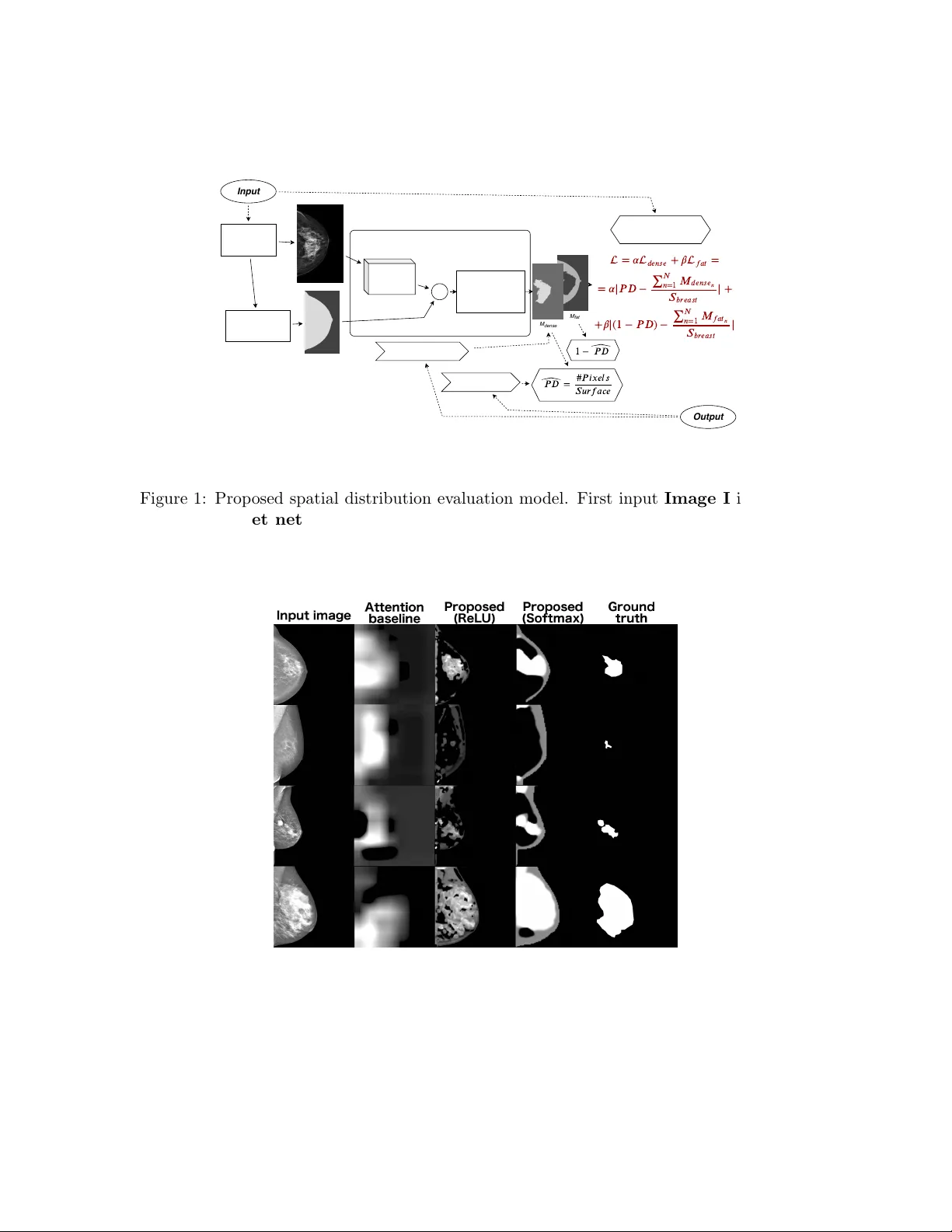
Medical Imaging with Deep Learning 2019 MIDL 2019 – Extended Abstract T rack A closer lo ok on to breast densit y with w eakly sup ervised dense-tissue masks Mic k ael T ardy 1 , 2 mickael.t ardy@ec-nantes.fr 1 LS2N UMR6004, Ec ole Centr ale Nantes, F r anc e, 2 Her a-MI, SAS, Nantes, F r anc e Bruno Sc heffer 2 , 3 bruno.scheffer@hera-mi.com 3 Institut de c anc´ er olo gie de l’Ouest, Saint-Herblain, F r anc e Diana Mateus 1 diana.ma teus@ec-nantes.fr Abstract This w ork fo cuses on the automatic quan tification of the breast density from digital mam- mograph y imaging. Using only categorical image-wise lab els w e train a mo del capable of predicting con tinuous density p ercentage as well as providing a pixel wise support frit for the dense region. In particular w e prop ose a weakly supervised loss linking the densit y p ercen tage to the mask size. Keyw ords: Deep Learning, W eakly supervised segmen tation, Regression, Breast density 1. Purp ose Breast densit y is a biomarker for breast cancer developmen t risk that suggests that the risk of cancer developmen t increases with denser breasts. Moreo ver, the detection of cancer in dense tissues and, more generally , in dense breasts is often considered more challenging due to the similar visual asp ects of normal and abnormal tissues, which complicates the in terpretation of mammographic images. F or the ab ov e reasons, we argue that computer- aided decision systems for early breast cancer detection should both, quan titatively ev aluate the breast density , and ev aluate the spatial distribution of the dense tissues. 2. Metho ds In clinical practice, breast densit y is usually assessed image-wise using a classification grid lik e the BI-RADS (Breast imaging-rep orting and data system) ( Irshad et al. , 2016 ). In the presen t work, w e prop ose to estimate breast densit y at the pixel level while using only image-wise ground truth from the BI-RADS scale. Our goal is to generate a breast densit y mask, iden tifying pixels asso ciated with the tissue that con tributed to the densit y class. T o achiev e our goal, we prop ose a nov el loss linking the sough t breast density mask to the globally estimated breast density (fig. 1 ). W e formulate the problem as a weakly sup ervised binary seman tic segmentation. Our approach is related to recen t efforts to reduce the amount of supervision ( Carneiro et al. , 2017 ; Dub ost et al. , 2017 ). In practice, w e rely on a mo dified U-Net architecture ( Ronneb erger et al. , 2015 ) and on an extended 12-class densit y grid that impro ves the density resolution compared to traditional BI-RADS classification (4th edition). Compared to the state-of-the-art, our c 2019 M. T ardy , B. Scheffer & D. Mateus. T ardy Scheffer Ma teus classification and segmen tation scheme do es not rely on the model ' s atten tion but uses a loss function efficien tly correlating a tissue mask with the target breast densit y v alues. Moreov er, the output is constrained with the breast binary mask removing useless activ ations. 3. Results The database for training and tests consists of 1232 and 370 images resp ectiv ely . W e got promising results with a mean absolute error (MAE) of 6.7% for the density regression estimate (see tab. 2 ) and an accuracy of 78% for 4-class BI-RADS density classification (tab. 1 ). Our comparison baseline is a V GG-like regression mo del trained on the same dataset. T o v alidate the segmen tation p erformance, we collected regions of interest on several images (16) and calculated the Dice = 0 . 65. Ov erall w e obtain clinically meaningful seg- men tation masks offering v aluable insigh ts in to the spatial distribution of the dense tissues (fig. 2 ). In comparison, w e demonstrate the inefficiency of the atten tion-based techniques for the breast density mask generation. In addition we v alidate our approach on the INBreast ( Moreira et al. , 2012 ) database. Without any additional training and a simple preprocessing w e obtained 65% accuracy and M AE = 13%. W e note that our results are comparable to other works on the same dataset (64 . 53%, ( Schebesch et al. ), 67 . 8% ( Angelo et al. , 2015 ). 4. Conclusions Our approac h to link breast densit y classification to the spatial distribution of dense tissue has a positive effect on classification scores while providing an additional output mask of the dense regions. These results are interesting given the considerably lo w requiremen ts on ground truth (just a class instead of an image mask) and the size of the training dataset. T able 1: 4-class BI-RADS classification p erformance. All mo dels are trained with 12-class grid. L is the prop osed loss. Metrics Model A c curacy Pr e cision R e call F 1 -sc or e Cohen kapp a VGG+Sigmoid+MSE 0.764 0.782 0.764 0.766 0.891 U-NET+Softmax+ L 0.684 0.729 0.684 0.679 0.838 U-NET+ReLU+ L 0.779 0.809 0.779 0.781 0.891 T able 2: Regression p erformances of the studied mo dels. All mo dels, except the last t wo are trained with 12-class grid. L is the prop osed loss. Metrics Dataset MAE ( % ) MxAE ( % ) C-index VGG+Sigmoid+MSE 6.545 31.964 0.820 U-NET+Softmax+ L 8.303 34.404 0.789 U-NET+ReLU+ L 6.661 32.156 0.839 2 A closer look onto breast density Breast density PD Neural Network Image I x Breast binary mask S breast Segmentation masks M U-Net Estimation Segmentation = + = = | − | + ∑ = 1 + | ( 1 − ) − | ∑ = 1 = ˆ # M dense M fat 1 − ˆ Input Output Figure 1: Prop osed spatial distribution ev aluation mo del. First input Image I is fed to a U-Net netw ork , then, u-net output is combined w ith a binary breast mask S breast to yield the output segmentation masks M = { M dense , M f at } . The com bined loss L guides the mo del training using image-wise P D density lab el. Figure 2: Resulting dense tissue masks. First column : input images, second column : activ ation masks pro duced by the atten tion-based baseline, third column : den- sit y masks M dense of ReLU-trained model, fourth column : density masks M dense of Softmax-trained mo del and fifth column : ground truth 3 T ardy Scheffer Ma teus Ac kno wledgments Researc h funding is pro vided b y Hera-MI, SAS and Asso ciation Nationale de la Recherc he et de la T ec hnologie via CIFRE grant no. 2018/0308. References Mic hele F ´ ulvia Angelo, P . C. Carneiro, T. C. Granado, and A. C. Patrocinio. Influ- ence of contrast enhancemen t to breast density classification by using sigmoid func- tion. In IFMBE Pr o c e e dings , volume 51, pages 33–36. Springer, Cham, 2015. ISBN 9783319193878. doi: 10.1007/978- 3- 319- 19387- 8 9. Gusta vo Carneiro, Tingying Peng, Christine Bay er, and Nassir Na v ab. Automatic Quan- tification of T umour Hyp o xia from Multi-Mo dal Microscop y Images Using W eakly- Sup ervised Learning Metho ds. IEEE T r ansactions on Me dic al Imaging , 36(7):1405–1417, jul 2017. ISSN 1558254X. doi: 10.1109/TMI.2017.2677479. Florian Dub ost, Gerda Bortso v a, Hieab Adams, Arfan Ikram, Wiro J Niessen, Meike V er- no oij, and Marleen De Bruijne. GP-Unet: Lesion Detection from W eak Lab els with a 3D Regression Net work. In Maxime Descoteaux, Lena Maier-Hein, Alfred F ranz, Pierre Jannin, D Louis Collins, and Simon Duchesne, editors, Me dic al Image Computing and Computer-Assiste d Intervention MICCAI 2017 , pages 214–221. Springer In ternational Publishing, 2017. ISBN 978-3-319-66179-7. Abid Irshad, Reb ecca Leddy , Susan Ack erman, Abbie Cluv er, Dag Pa vic, Ahad Abid, and Madelene C. Lewis. Effects of changes in BI-RADS densit y assessment guidelines (fourth v ersus fifth edition) on breast density assessmen t: In tra-and in terreader agreemen ts and densit y distribution. Americ an Journal of R o entgenolo gy , 207(6):1366–1371, dec 2016. ISSN 15463141. doi: 10.2214/AJR.16.16561. In ˆ es C. Moreira, Igor Amaral, In ˆ es Domingues, Ant´ onio Cardoso, Maria Jo˜ ao Cardoso, and Jaime S. Cardoso. INbreast: T ow ard a F ull-field Digital Mammographic Database. A c ademic R adiolo gy , 19(2):236–248, 2012. ISSN 10766332. doi: 10.1016/j.acra.2011.09. 014. Olaf Ronneb erger, Philipp Fischer, and Thomas Bro x. U-net: Conv olutional net w orks for biomedical image segmentation. T ec hnical rep ort, 2015. F rank Sc heb esc h, Mathias Unberath, Ingwer Andersen, and Andreas Maier. Breast den- sit y assessment using wa velet features on mammograms. In Informatik aktuel l , pages 38–43. Springer View eg, Berlin, Heidelb erg. ISBN 9783662494646. doi: 10.1007/ 978- 3- 662- 49465- 3 9. 4
Original Paper
Loading high-quality paper...
Comments & Academic Discussion
Loading comments...
Leave a Comment