In this paper, we studied the association between the change of structural brain volumes to the potential development of Alzheimer's disease (AD). Using a simple abstraction technique, we converted regional cortical and subcortical volume differences over two time points for each study subject into a graph. We then obtained substructures of interest using a graph decomposition algorithm in order to extract pivotal nodes via multi-view feature selection. Intensive experiments using robust classification frameworks were conducted to evaluate the performance of using the brain substructures obtained under different thresholds. The results indicated that compact substructures acquired by examining the differences between patient groups were sufficient to discriminate between AD and healthy controls with an area under the receiver operating curve of 0.72.
Deep Dive into From Brain Imaging to Graph Analysis: a study on ADNIs patient cohort.
In this paper, we studied the association between the change of structural brain volumes to the potential development of Alzheimer’s disease (AD). Using a simple abstraction technique, we converted regional cortical and subcortical volume differences over two time points for each study subject into a graph. We then obtained substructures of interest using a graph decomposition algorithm in order to extract pivotal nodes via multi-view feature selection. Intensive experiments using robust classification frameworks were conducted to evaluate the performance of using the brain substructures obtained under different thresholds. The results indicated that compact substructures acquired by examining the differences between patient groups were sufficient to discriminate between AD and healthy controls with an area under the receiver operating curve of 0.72.
From Brain Imaging to Graph Analysis: a study on ADNI’s patient cohort
Rui Zhang, PhD1, Luca Giancardo2, Danilo A. Pena2, Yejin Kim2,
Hanghang Tong1, Xiaoqian Jiang2; for the Alzheimer's Disease Neuroimaging Initiative*
1School of Computing, Infor. & Decis. Syst. Engin., Arizona State University
2School of Biomedical Informatics, University of Texas Health Science Center at Houston
ABSTRACT
In this paper, we studied the association between the change of structural brain volumes to the potential
development of Alzheimer’s disease (AD). Using a simple abstraction technique, we converted regional cortical and
subcortical volume differences over two time points for each study subject into a graph. We then obtained
substructures of interest using a graph decomposition algorithm in order to extract pivotal nodes via multi-view
feature selection. Intensive experiments using robust classification frameworks were conducted to evaluate the
performance of using the brain substructures obtained under different thresholds. The results indicated that compact
substructures acquired by examining the differences between patient groups were sufficient to discriminate between
AD and healthy controls with an area under the receiver operating curve of 0.72.
INTRODUCTION
Brain functionality and decreasing cognition is known to be associated with the progression of Alzheimer’s disease
(AD). The difference between asymptomatic and symptomatic AD can change over time, and this period between
being a healthy individual to having clinically present AD is referred to as mild cognitive impairment (MCI). This
functional and cognitive decline is marked with memory lapses, poorer executive function, and increasing
complexities associated with common activities of daily living that can last years [1]. The standard diagnosis of AD
patients typically begins with a series of neuropsychological tests, clinical assessments, followed by various imaging
tests. Over time, patients undergo multiple brain imaging visits at different points which allow physicians to link
physical manifestations of AD to lab and clinical measurement data. This wealth of information enables researchers
to take advantage of these multi-visit data to look at the AD neurodegeneration process (through observing cognitive
decline) both cross-sectionally and longitudinally. Recent studies have used many methods look at AD from a
different perspective. Researchers use techniques such as hierarchical classification [2], convolutional neural
networks [3], tract-based spatial statistics [4], in addition to a whole series of multi-modality data [5] to improve AD
diagnostic performance. Further, studies have looked at various combinations of phenotype classification, typically
incorporating both stable and converted MCI patients [6]. This allows for a rich understanding of AD progression
and of biomarkers that may be readily available to drive an improved and quicker AD diagnosis. In this work, we
aim to leverage the advantages of graph-based approaches while discarding some of the disadvantages that come
with diffusion MRI based techniques. Specifically, we used structural volume data to uncover how different regions
of the brain are inter-connected during AD progression. We also incorporated longitudinal information which
accounts for structural changes in a patient’s brain over time. These inter-region network effects will be used for
phenotype classification to demonstrate their discrimination power.
RELATED WORK
Graph theory and graph-based approaches have been studied for many years in the neuroimaging community. These
networks have been created using several different modalities including structural magnetic resonance imaging
(MRI), diffusion tensor imaging (DTI), and positron emission tomography (PET) for various diseases such as AD.
Typically, these approaches look at the differences between normal controls (CN) and those with the studied
disease. The subsequent analysis allows for interpretation of nodes, hubs, a
…(Full text truncated)…
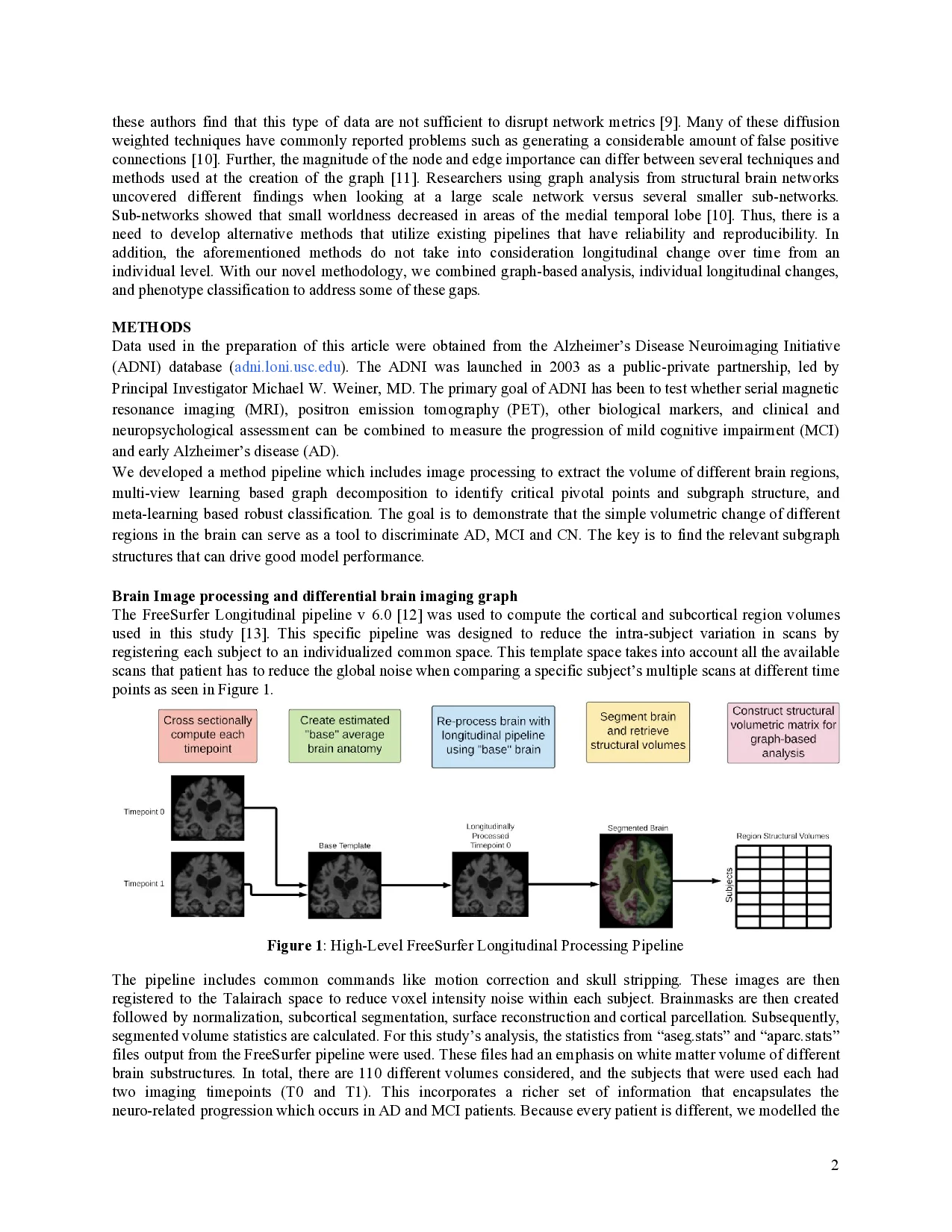
This content is AI-processed based on ArXiv data.