Prior Information Guided Regularized Deep Learning for Cell Nucleus Detection
Cell nuclei detection is a challenging research topic because of limitations in cellular image quality and diversity of nuclear morphology, i.e. varying nuclei shapes, sizes, and overlaps between multiple cell nuclei. This has been a topic of endurin…
Authors: Mohammad Tofighi, Tiantong Guo, Jairam K.P. Vanamala
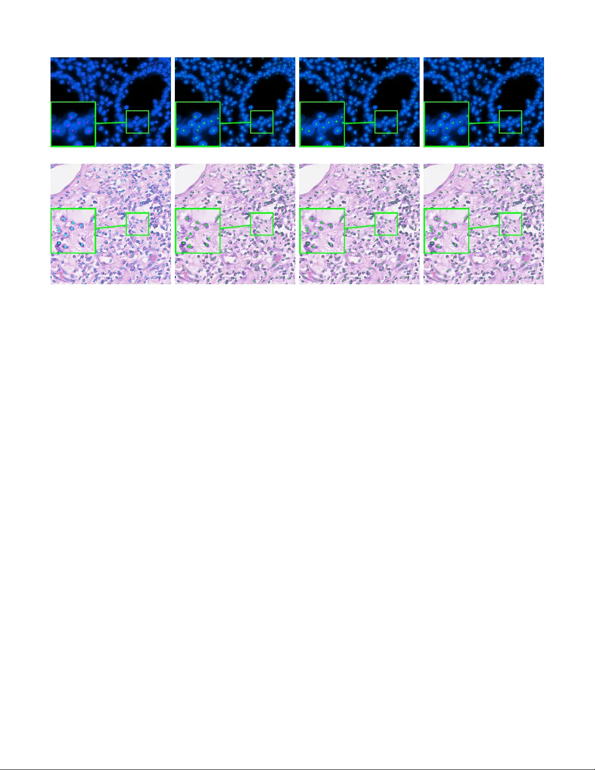
IEEE TRANSA CTIONS ON MEDICAL IMA GING, A CCEPTED FOR PUBLICA TION, J ANU AR Y 2019 1 Prior Information Guided Re gularized Deep Learning for Cell Nucleus Detection Mohammad T ofighi, Student Member , IEEE, T iantong Guo, Student Member , IEEE, Jairam K.P . V anamala, and V ishal Monga, Senior Member , IEEE Abstract —Cell nuclei detection is a challenging r esearch topic because of limitations in cellular image quality and diversity of nuclear morphology , i.e. varying nuclei shapes, sizes, and overlaps between multiple cell nuclei. This has been a topic of enduring interest with promising recent success shown by deep learning methods. These methods train Convolutional Neural Networks (CNNs) with a training set of input images and known, labeled nuclei locations. Many such methods are supplemented by spatial or morphological pr ocessing. Using a set of canonical cell nuclei shapes, prepared with the help of a domain expert, we develop a new appr oach that we call Shape Priors with Con v olutional Neural Networks (SP-CNN). W e further extend the network to introduce a shape prior (SP) layer and then allowing it to become trainable (i.e. optimizable). W e call this network tunable SP-CNN (TSP-CNN). In summary , we present new network structur es that can incorporate ‘expected beha vior’ of nucleus shapes via two components: learnable layers that perform the nucleus detection and a fixed processing part that guides the learning with prior information. Analytically , we formulate two new regularization terms that are targeted at: 1) lear ning the shapes, 2) reducing false positives while simultaneously encouraging detection inside the cell nucleus boundary . Experimental results on two challeng- ing datasets reveal that the proposed SP-CNN and TSP-CNN can outperform state-of-the-art alternativ es. Index T erms —nucleus detection, deep learning, conv olutional neural networks, shape priors, learnable shapes I . I N T RO D U C T I O N Microscopic images often exhibit high degree of cellular heterogeneity [1], [2]. Cell nucleus detection methods locate the cell and annotate the center of the cell nuclei. V isual-based techniques in many medical imaging applications, e.g. manual detection and counting, are extremely time consuming and do not scale well as the task is to be performed for a large number of images [3], [4]. Therefore, automatic analysis of cellular imagery to determine nuclei locations is a centrally important problem in diagnosis of se veral medical conditions including tumor and cancer detection [5], [6]. Related work: Some of the earliest attempts at cell nuclei detection in v olved tailored feature extraction and morpholog- ical processing [7], [8], [9], [10]. One key limitation of these approaches is that the best features for nuclei detection are rarely readily apparent. Further , often the designed techniques are too specific to the choice of dataset and not versatile. Because of their ability to perform feature discovery and M. T ofighi, T . Guo, and V . Monga are with the Department of Electrical Engineering, Pennsylv ania State University , Uni versity Park, P A. e-mail: tofighi@psu.edu, tiantong@psu.edu, juv4@psu.edu, vmonga@engr .psu.edu. J.K.P . V anamala is with Center for Molecular Immunology and Infectious Disease, Pennsylvania State University , University Park, P A. Research has been supported by an NSF CAREER award (to V . Monga). generalizable inference, deep learning methods hav e recently become popular for this problem [11], [12], [13], [14]. For instance, Cruz-Roa et al. [15] showed that a deep learning architecture for nuclei detection outperforms methods based on different image representation strategies e.g. bag of features, canonical and wav elet transforms. Xie et al. [16], proposed a structural regression model for CNN, where a cell nuclei center is detected if it has the maximum value in the proximity map. In [17], Xu et al. proposed a cell detection method based on a stacked sparse autoencoder, where it learns high lev el features of cell centroids and then a softmax classifier is used to separate the nuclear and non-nuclear image patches. Sirinukunwattana et al. proposed SC-CNN [5] which uses a regression approach to find the likelihood of a pixel being the center of a nucleus. In SC-CNN, the probability values are topologically constrained in a way that in vicinity of nuclei center the probability is higher . Another recent approach [18] uses a combination of well-known traditional CNNs for cell nuclei segmentation and dictionary learning techniques for refining segmentations results. Combining CNNs with sparse coding, [19] uses a sparse con v olutional autoencoder (CSP-CNN) for simultaneous nucleus detection and feature extraction. In CSP-CNN, the detection is based on a fairly deep network ( > 15 layers) comprising of multiple CNN branches that perform detection, se gmentation and image reconstruction. W e de velop a novel prior guided learning approach by exploiting a prior understanding of the shape of the nuclei, which can be obtained in consultation with a domain expert. The value of shape based information has been recognized for medical image segmentation [20], [21]. Le venton et al. [22] proposed a method for medical image segmentation by incorporating shape information into the geodesic active contour method. Ali et al. [23] incorporated prior shape information into boundary and region based activ e contours for accurate segmentation of cells. In [24], Oktay et al. propose to incorporate shape prior information into enhancement and segmentation of MR cardiac images. They propose a fidelity term between the output segmentation regions and the ground- truth region shapes. While the approach in [24] also exploits anatomical shape information, technically the proposed SP- CNN is fundamentally dif ferent. First, the image modality in our work is cellular as opposed to org an (MR) imagery in [24], which leads to vastly different quantitative formulations. Second, our proposed regularizer is not a fidelity term but guides cell nuclei detection in regions where the probability of presence of a cell nuclei is higher according to the shape prior knowledge. Finally , architecturally the proposed SP/TSP- IEEE TRANSA CTIONS ON MEDICAL IMA GING, A CCEPTED FOR PUBLICA TION, J ANU AR Y 2019 2 CNN and the neural network in [24] are entirely different. Prior information with deep networks has emerged as a promising direction in imaging inv erse problems such as super-resolution [25], [26]. These methods use output image priors to enhance the super-resolution task. Similarly in related work, a Deep Image Prior [27] network has been specifically designed for tasks such as image in-painting, de-noising, and super-resolution. Shape based priors in a deep learning based nuclei detection framework ho wev er remain elusiv e and form the focus of our work. Contributions: While existing deep learning approaches for cell nuclei detection are promising, our goal is to fundamen- tally alter the learning of the network by enriching it with domain knowledge provided by a medical expert. Specifically , our ke y contributions 1 are as follo ws: • Shape Prior with a Con volutional Neural Network (SP-CNN): W e propose a novel network structure that can incorporate ‘expected behavior’ of nuclei shapes via two components: learnable layers that perform the nu- cleus detection and a fixed processing part that guides the learning with prior information. Three sources contribute to generating the prior: network output, raw edge map from the input image, and a set of predefined shapes prepared by the medical expert. These shapes are binary images representing the boundary of cell nuclei. The fixed priors guide the learnable layers to perform detection consistent with a nucleus boundary . • T unable Shape Priors with a Con v olutional Neural Network (TSP-CNN): As a key extension, we dev elop a new approach where the set of shapes is represented as a con v olutional layer , which is no-longer fixed but learned. That is, using the expert provided shapes as a starting point, we both refine and remove redundancy from the shape set (using shape similarity measures) to arriv e at a new learned shape set that is not only more economical but also enhances detection accuracy . • Novel Regularization T erms: Analytically , we formulate two new regularization terms: a shape prior term and a shape learning term. The shape prior term is used in both SP-CNN and TSP-CNN and incorporates the effect of prior information by using a set of shapes to guide the learning of the network towards greater accuracy . The shape learning term is only used in TSP- CNN and achieves the refinement/learning of shapes in a constrained manner, i.e. by emphasizing similarity to a reference shape set. W e carefully design these terms so they are differentiable w .r .t. the output and hence the network parameters, enabling tractable learning through standard back-propagation schemes. • A New Dataset, Broad Experimental V alidation and Reproducibility: Experimental v alidation of SP-CNN and TSP-CNN is carried out on two div erse cancer tissue datasets to show its broad applicability . W e also provide a new open source expert annotated dataset of microscopic colon tissue images for cell nuclei detection. 1 A preliminary 4 page version of this work was published at IEEE ICIP 2018 in Athens, Greece held in October 2018 [28]. Fig. 1: Samples of handcrafted cell nuclei shapes from colon tissues in UW Dataset. For dataset details see Sec. IV. W e call it the PSU Dataset and it is prepared by the help of experts in Center for Molecular Immunology and Infectious Disease, Penn State Uni versity [29]. The second dataset is courtesy of Sirinukunwattana et al. at Department of Computer Science, Univ ersity of W arwick (we call it UW Dataset) which includes colorectal adeno- carcinoma images [5]. Extensive experimental results in the form of benchmark measures such as F1 scores and precision-recall curves show that SP-CNN and TSP-CNN outperform many recent methods [5], [16], [7], [17], [30], [19], particularly in very challenging scenarios. W e also make our code and the PSU dataset freely available at our project webpage [29]. The rest of this paper is organized as follows. The pro- posed SP-CNN structure that incorporates shape priors into cell nuclei detection and formulation of the ne w re gularized cost function are detailed in Section II. Section III presents the extension of SP-CNN to tunable shape priors. Detailed experimental comparisons against competing state-of-the-art methods are provided in Section IV. Section V summarizes the findings and concludes the paper . I I . S H A P E P R I O R S W I T H C O N VO L U T I O N A L N E U R A L N E T W O R K S ( S P - C N N ) A. CNN for Nucleus Detection SP-CNN detects the cell nucleus using a regression CNN. In regression networks, the goal is to obtain a function that interprets the relationship between the input image x and ground-truth labeled image y . The network is modeled by parameters set Θ = { W l , b l } D l =1 , where W l and b l denote respectiv ely the weights and bias of l -th layer of total D layers collectiv ely . The CNN is learned by solving the well-kno wn optimization problem [5], [31]: Θ = arg min Θ k f ( x ; Θ ) − y k 2 2 (1) where f ( x ; Θ ) represents the non-linear mapping of the CNN that generates the detection maps ˆ y . W e work with soft labels y and ˆ y , i.e. y , ˆ y can take values in the range [0 , 1] . In practice, y is obtained by processing the binary image (0 or 1 at each pixel) of ground-truth nuclei locations as in [5]; also see Section IV. Each of the D CNN layers comprises of a con v olutional layer follo wed by an activ ation function, which is a Rectified Linear Unit (ReLU) [32] (except the last layer). B. Deep Networks W ith Shape Priors As discussed in Section I, we now incorporate the prior information about the shape of the cell nuclei into the training IEEE TRANSA CTIONS ON MEDICAL IMA GING, A CCEPTED FOR PUBLICA TION, J ANU AR Y 2019 3 Prio r Info rmation F unctionalit y Raw Edges ˆ x Edge Map Masked g p ( ˆ y ) ˆ x { S i } p rio rs Window Map g p ( ˆ y ) Raw Input x ...... Output ˆ y P Prior Information D -la y er CNN { Θ } Generate Raw Edges Generate Windo w Map Element-wise multiplication Influencing CNN Fig. 2: SP-CNN illustration. There are two parts of the SP-CNN: Functionality part (blue) and Prior Information part (orange). The Functionality part consists of one D -layer CNN that takes input raw image x and generates the detected labels for cell nuclei ˆ y . The prior information part computes the prior cost term as in Eq. (2) and feeds the information into the (learning of the) CNN through back-propagation to guide it towards enhanced nuclei detection. Note the prior information part generates a regularization term used in training the D learnable layers of the CNN and only the functionality part of the network is applied to a test image. Hence, while the black, orange, and blue arrows are used during training, in testing stage only the black arro ws are used. of the CNN. Ideally , the labels produced by the CNN should lie inside of the nuclei boundaries. W e set up a regularization term to explicitly encourage the learned network to achieve detection inside the nucleus boundary while simultaneously reducing false positives. The regularizer is based on a set of shape priors developed with the help of domain expert and giv en by: S = {S i | i = 1 , 2 , . . . , N } . For each dataset, multiple training images are analyzed by a medical expert to hand label the nuclei boundaries. A set of N representativ e shapes is then hand crafted by the medical expert to form the set S . Some examples of the nucleus shape priors are shown in Fig. 1. These are corresponding to colon tissue images – detailed explanation is provided in Section IV. T o construct a meaningful regularization term emphasizing shape priors, we need the nucleus boundary information of the input raw image x . W e employ the widely used Canny edge detection 2 filter [33] to generate the raw edge image ˆ x with edges labeled as 1 and background as 0 , as shown in Fig. 3- (b). Note that the raw edge image ˆ x is only used during the training process. W e no w define the regularization term that captures shape priors: L SP = − λ N X i =1 k ( g p ( ˆ y ) ˆ x ) ∗ S i k 2 2 (2) where the term g p ( · ) denotes the max pooling operation on ˆ y with window size p , represents element-wise multiplication, and ∗ is the 2D con v olution operation. Based on Eq. (2), the computation of the shape priors cost term consists of three 2 Note we select Canny edge detection filter because of its simplicity and efficiency . Although its performance satisfies our intentions, other edge detection methods can also be used. (a) Raw input ( x ) (b) Raw edges ( ˆ x ) (c) Window map g p ( ˆ y ) (d) Masked edges ( g p ( ˆ y ) ˆ x ) (e) Output ( ˆ y ) (f ) T rue lab els ( y ) (a) Ra w input ( x ) (b) Ra w edges ( ˆ x ) (c) Windo w map g p ( ˆ y ) (d) Mask ed edges ( g p ( ˆ y ) ˆ x ) (e) Output ( ˆ y ) (f ) T rue lab els ( y ) (a) Ra w input ( x ) (b) Ra w edges ( ˆ x ) (c) Windo w map g p ( ˆ y ) (d) Mask ed edges ( g p ( ˆ y ) ˆ x ) (e) Output ( ˆ y ) (f ) T rue lab els ( y ) (a) Ra w input ( x ) (b) Ra w edges ( ˆ x ) (c) Windo w map g p ( ˆ y ) (d) Mask ed edges ( g p ( ˆ y ) ˆ x ) (e) Output ( ˆ y ) (f ) T rue lab els ( y ) (a) Ra w input ( x ) (b) Ra w edges ( ˆ x ) (c) Windo w map g p ( ˆ y ) (d) Mask ed edges ( g p ( ˆ y ) ˆ x ) (e) Output ( ˆ y ) (f ) T rue lab els ( y ) (a) Ra w input ( x ) (b) Ra w edges ( ˆ x ) (c) Windo w map g p ( ˆ y ) (d) Mask ed edges ( g p ( ˆ y ) ˆ x ) (e) Output ( ˆ y ) (f ) T rue lab els ( y ) (a) Ra w input ( x ) (b) Ra w edges ( ˆ x ) (c) Windo w map g p ( ˆ y ) (d) Mask ed edges ( g p ( ˆ y ) ˆ x ) (e) Output ( ˆ y ) (f ) T rue lab els ( y ) Fig. 3: Images in each step of SP-CNN. steps as sho wn in Fig. 2’ s prior information (orange) part: 1) The CNN output ˆ y is first thresholded by T p = 0 . 2 to eliminate the background noise (Fig. 3-(e)) and then max pooled by g p ( · ) with stride of 1 and the ‘SAME’ padding scheme 3 . This results in a window map g p ( ˆ y ) that has p × p window centered at each location within the soft detected region (Fig. 3-(c)). As expected, a window with higher numerical v alue will result if the detected label values (in ˆ y ) are correspondingly higher (closer to 1). 2) The window map g p ( ˆ y ) is then multiplied with the raw edge image ˆ x element-wise. This step serves to mask 3 Because there is no change in the image grid, this is similar to the box- filtering operation in image processing. IEEE TRANSA CTIONS ON MEDICAL IMA GING, A CCEPTED FOR PUBLICA TION, J ANU AR Y 2019 4 out the edges from ˆ x that surround the detected location in ˆ y , as shown in Fig. 3-(d). 3) The masked edge image ( g p ( ˆ y ) ˆ x ) is con v olved with the shape priors in set S to generate a measurement of ho w well does the detection fit inside the nucleus shape. If ˆ y has more labels predicted inside the nucleus boundary , Eq. (2) will achie ve a smaller value (more negati v e). Note that the effect of the shape prior is captured by a negati v e regularization term since the goal is to maximize (and not minimize) correlation with ‘expected shapes’. Overall, the cost function of the SP-CNN is given by: Θ = arg min Θ L Loss + L SP (3) = arg min Θ k f ( x ; Θ ) − y k 2 2 − λ N X i =1 k ( g p ( ˆ y ) ˆ x ) ∗ S i k 2 2 where λ is the trade-off parameter between the squared loss term and the regularizer representing the effect of the shape prior . The term L SP is carefully designed to simultaneously accomplish two tasks: 1.) a high detection rate of nuclei is en- couraged since the element-wise Hadamard product highlights edge boundaries, and 2.) the subsequent conv olution with the expert provided shape set reduces false positives. Note that ˆ y := f ( x ; Θ ) , thus the shape prior cost term is effected by the network parameters and also introduces gradient terms that updates the network parameters during the training process using back-propagation [34]. This is indicated in Fig. 2 by dashed line under “Influencing CNN”. Note that while the black, orange, and blue arro ws are used during training, the black arro ws are just used in testing stage. Let L = L Loss + L SP be the cost function. At iteration t of the back-propagation, the filters of CNN are updated as: Θ t +1 = Θ t − η ∇ Θ L (4) where η denotes the learning rate, and Θ = { W k , b k } D k =1 . The follo wing gradients are to be computed 4 : ∂ L ∂ W k , ∂ L ∂ b k where W k and b k denote one of the filters and biases at k -th layer of the CNN, representatively . The equation for computing the gradient w .r .t. an arbitrary entry within filter W k in layer k ∈ { 1 , ..., D } is given by: ∂ L ∂ W a k = − < ( ˆ y − y ) , ∂ ˆ y ∂ W a k > F (5) − λ N X i =1 < ( g p ( ˆ y ) ˆ x ) ∗ S i , g − 1 p ∂ ˆ y ∂ W a k ˆ x ∗ S i > F where W a k denotes an arbitrary scalar entry within the repre- sentativ e filter W k , < · , · > F denotes the real value Frobenius inner product 5 , and g − 1 p ( · ) assigns the deriv ati v e to where 4 Note the update rules and the gradients for the bias terms are similar and are included with more details in the supplementary document [29]. 5 For two real valued matrices A and B with same dimension, < A , B > F := P i,j A i,j B i,j where i, j are the indexes of the entries. activ ations come from, the “winning unit”, because other units in the previous layer’ s pooling blocks did not contribute to it. Hence deri v ativ e of all the other units is assigned to e zero. In Eq. (5), ∂ y ∂ W a k is computed by following the standard backpropagation rule for each layer k [35]. I I I . T U NA B L E S H A P E P R I O R S W I T H C O N VO L U T I O N A L N E U R A L N E T W O R K S ( T S P - C N N ) While exploiting a shape set provided by a domain expert is meritorious, a fundamental open question is: What is the best, most compact shape set to employ and can it be adapted based on the image characteristics? This section provides an answer to that question in the following manner: the set of shapes can be seen as a collection of filters, which we now add to the network as a learnable component and call it the learnable shape layer – see Fig. 4 for an illustration of this idea. W e call this new network structure as T unable Shape Priors with CNNs (TSP-CNN). W e observe that the domain expert provided shape set in practice is quite redundant, i.e. it may contain pairs of hand- crafted shapes that are quite close to each other and do not add value to nucleus detection. W e therefore propose a two stage process to arri ve at a compact Basis Shape Set which is adapted based on training imagery: 1.) shape elimination stage to obtain a reduced/smaller reference shape set, and 2.) shape refinement or learning via TSP-CNN. This two stage process is illustrated in Fig. 5. For both stages, meaningful shape similarity measures are needed; the exact shape elim- ination and refinement procedure is detailed ne xt. W e next introduce shape elimination and refinement procedures, and then describe the training procedure to learn shapes that are adapted to the underlying dataset. A. Shape Elimination Many shape similarity measures hav e been dev eloped in image processing and vision for a v ariety of different tasks [36], [37], [38], [39], [40], [41], [42]. Most of these methods measure the distance between shapes A and B , d ( A, B ) in different ways, for example bottleneck distance, Hausdroff distance, turning function distance, etc [36]. Attalla et al. used a multi-resolutional polygonal shape descriptor that is in v ariant to scale and is used to match and recognize 2D shapes. In [39], Latecki et al. proposed a cognitiv ely motiv ated similarity measure which divides a shape into best possible correspondences of visual parts and measures the similarity between each of them and then aggre gates them to find the ov erall similarity measure. In [37], Sampat et al. proposed Complex W avelet SSIM (CW -SSIM) which is an extension of the well-known Struc- tural Similarity Index Measure (SSIM) [43]. CW -SSIM has been shown to be insensitiv e to nonstructural geometric dis- tortions and an e xcellent measure to compare binary shape similarity [37] because of its ability to capture phase infor- mation. CW -SSIM index [37] for two sets of coef ficients in the complex wa velet transform domain as c x = { c x,i | i = 1 , . . . , M } and c y = { c y ,i | i = 1 , . . . , M } , extracted from IEEE TRANSA CTIONS ON MEDICAL IMA GING, A CCEPTED FOR PUBLICA TION, J ANU AR Y 2019 5 Prio r Info rmation F unctionalit y Raw Edges ˆ x Window Map Masked g p ( ˆ y ) ˆ x ... ... Window Map g p ( ˆ y ) Raw Input x ...... Output ˆ y Learnable { S l i } P D -la y er CNN { Θ } Generate Raw Edges Generate Windo w Map Element-wise multiplication Influencing CNN Fig. 4: TSP-CNN illustration. There are two parts of TSP-CNN: Functionality part and Prior Information part. Compared to SP-CNN, the shape set S l is no longer fixed. It is now also a learnable component of the network. The shapes in the shape set are updated according to the shape learning regularization term as in Eq. (8). As with SP-CNN, the prior information part generates regularization terms used in training the D learnable layers of the CNN (through the blue and orange arrows) but only the functionality part of the network, i.e. the D layer CNN (excluding the learned shapes) is applied to a test image to predict nuclei centers (through the black arrows). same location of same wav elet subbands of two shape images being compared is defined as: CW -SSIM ( c x , c y ) = 2 | P M i =1 c x,i c ∗ y ,i | + K P M i =1 | c x,i | 2 + P M i =1 | c y ,i | 2 + K , (6) where c ∗ is the complex conjugate of c and K is a small positiv e constant. After computing CW -SSIM values for all sets, c i ’ s, the final CW -SSIM value for two shape images CW -SSIM ( S i , S j ) is obtained by av eraging CW -SSIM value ov er all wavelet subbands as described in [37]. Starting with the domain expert provided shape set S = {S i | i = 1 , 2 , . . . , N } , we eliminate redundancy by using the aforementioned CW -SSIM measure to compute pairwise comparisons of shapes. Near identical (redundant) shapes are grouped together and one representativ e is extracted from the group. The complete procedure is detailed in Algorithm 1. B. Shape Refinement The output of Algorithm 1 is what we address as S r = {S r j | j = 1 , 2 , . . . , Q } the Reference Shape Set – which is essentially a cardinality (size) reduced version of the domain expert provided shape set S , i.e. Q < N . Our approach to learn or refine shapes is then to optimize a Learnable Shape Set S l = {S l i | i = 1 , 2 , . . . , Q } under the important physical constraint that the shapes in S l bear similarity to those in S r . Ideally , as in the shape elimination stage, CW -SSIM should be used as a similarity measure between S l and S r . Howe ver , 6 In Algorithm 1, {} is an empty set, | S | is the cardinality of set S , and Rep ( S n ) is one representative shape from grouped similar shapes in set S n . Algorithm 1 Shape elimination procedure using CW -SSIM Input: S = {S i | i = 1 , 2 , . . . , N } , k , n = 1 , b S , S n = {} 6 1: while k = 1 do 2: S n ← S n ∪ {S k } 3: for l = 2 to N do 4: C = CW -SSIM ( S k , S l ) 5: if C > 0 . 8 then 6: S n ← S n ∪ {S l } 7: else 8: Go to line 3 9: end if 10: end f or 11: S ← S \ { S ∩ S n } 12: b S ← b S ∪ { Rep ( S n ) } 13: n ← n + 1 14: N = | S | 15: if N = 0 then 16: k = 0 17: end if 18: end while Output: Reference shape set S r = b S . CW -SSIM is not differentiable and hence arduous to incor- porate in standard CNN learning framew orks which rely on deriv ati v e based back-propagation. SSIM, a pre-cursor to CW - SSIM on the other hand is dif ferentiable and implementable in a deep learning frame work [44]. In Fig. 6, we compare CW -SSIM and SSIM v alues in four cases of binary shape IEEE TRANSA CTIONS ON MEDICAL IMA GING, A CCEPTED FOR PUBLICA TION, J ANU AR Y 2019 6 ... ... ... Shap e Elimination Shap e Refinement A B C Fig. 5: An illustration of the two-stage process of shape elimination and refinement. Starting with the domain expert provided shape set, a redundancy removing elimination step is performed first using Algorithm 1. Second, refinement of this reduced size shape set is performed which adapts to training imagery and arri ves at a compact Basis Shape Set . Case-1 Case-2 Case-3 Case-4 Case-5 0 0 . 2 0 . 4 0 . 6 0 . 8 1 0 . 85 0 . 72 0 . 53 0 . 18 0 . 54 0 . 78 0 . 56 0 . 32 0 . 21 0 . 39 CW-SSIM SSIM , , , , , Fig. 6: SSIM & CW -SSIM values for different cases of shapes similarity by using representativ e nuclei shapes corresponding to the image datasets we work with: case-1: very similar , case-2: similar , case-3: different, case-4: very different, and case-5: rotation ( 90 ◦ ). From Fig. 6 we can infer two facts: 1.) CW -SSIM can be quite effecti ve in comparing binary nuclei shapes, and 2) SSIM forms an approximation to CW -SSIM. Giv en the tractability benefits of SSIM in deep learning set- ups, we employ it to build our shape similarity regularizer . As is corroborated later in Section IV, using an SSIM based regularizer suffices to afford TSP-CNN significant practical gains. The design of more sophisticated e.g. rotation in v ariant, similarity measures that can be tractably incorporated in deep learning frame works is a viable direction for future research. The SSIM index between patches x ∈ S l i and y ∈ S r j is SSIM ( x , y ) = 2 µ x µ y + C 1 µ 2 x + µ 2 y + C 1 . 2 σ xy + C 2 σ 2 x + σ 2 y + C 2 , (7) where µ and σ are mean and standard deviation of the patches, respectiv ely . The patch selection, comparisons and estimation of local statistics is done as in [43]. The overall SSIM v alue between two shapes, SSIM ( S l i , S r j ) , is the av erage of SSIM value ov er all the patches in the shapes. In [44], the use of SSIM is moti vated by image quality concerns. In TSP-CNN, dif ferently from [44], we use SSIM to define a shape learning regularizer in the following manner: L SSIM = − γ SSIM ( S l , S r ) = − γ Q X i =1 Q X j =1 SSIM ( S l i , S r j ) (8) and optimize the network parameters (including shapes) with: b Θ = arg min b Θ L Loss + L SP + L SSIM = arg min b Θ k f ( x ; b Θ ) − y k 2 2 − λ Q X i =1 k ( g p ( ˆ y ) ˆ x ) ∗ S l i k 2 2 − γ Q X i =1 Q X j =1 SSIM ( S l i , S r j ) (9) where b Θ = { Θ , S l } = { W , b , S l } . In Eq. 9, L Loss is the standard mean squared loss term between the ground-truth and output of the network. Note that reference shape set S r is obtained using the Algorithm 1, that is S r is a pruned shape set obtained using the shape elimination procedure. The shapes that are actually learned are in the set S l , and therefore S l once optimized represents the refined version of the shapes in S r . The shapes in set S l are learned according to the shape prior term ( L SP ) and the shape similarity measure ( L SSIM ): in particular , as argued before the shape prior term L SP should be minimized – so in essence, we are looking for the best shape set S l that can do so. Note that the second regularizer L SSIM emphasizes similarity to the pruned reference shape set S r and therefore ensures that physically meaningful shapes are learned – unconstrained optimization of S l could lead to learned shapes that do not conform to reality . C. Back-pr opagation: Shape Learning Cost T erm The back-propagation for TSP-CNN is similar to that of SP-CNN, except for the new shape learning term. The back- propagation for shape learning term only depends on the shapes S l i . Let the cost function be b L = L Loss + L SP + L SSIM , hence its gradient w .r .t. S a l i , an arbitrary scalar entry within the shape S l i , is as follows: ∂ b L ∂ S a l i = − λ Q X i =1 < ( g p ( ˆ y ) ˆ x ) ∗ S l i , ( g p ( ˆ y ) ˆ x ) ∗ I > F − γ Q X j =1 ∂ ∂ S a l i SSIM ( S l i , S r j ) (10) where I denotes a matrix with same size as S l i with only entrance a active as one. Second term in right hand side of Eq. 10 is derived in detail in [45], [44]. For a complete deriv ation of the back-propagation procedure, refer to our supplementary document in [29]. IEEE TRANSA CTIONS ON MEDICAL IMA GING, A CCEPTED FOR PUBLICA TION, J ANU AR Y 2019 7 PSU Dataset ( 30 × 30 ) UW Dataset ( 20 × 20 ) Fig. 7: Learned basis shapes using TSP-CNN for both UW and PSU datasets. Note that our optimization of shapes does not constrain them to be binary and hence these shapes have thicker boundaries with values in between 0 and 1. I V . E X P E R I M E N TA L R E S U LT S A. Experimental Setup and Datasets W e train and test SP-CNN and TSP-CNN on two colon cancer datasets: 1) publicly av ailable UW Dataset [5] which includes 100 H&E stained histology images of colorectal adenocarcinomas. There are a total number of 29756 nuclei marked at the nucleus center (please refer to Sec. VII.A. of [5] for more information). Our choice of the UW Dataset is because it represents real-world challenges such as o verlapping nuclei and contains other shapes that are often confused with nuclei. Further, it is one of the few publicly av ailable datasets that is widely used in many recent deep learning-based nuclei detection methods [5], [19], [46], 2) a new dataset carefully prepared and labeled manually by medical experts at the Cen- ter for Molecular Immunology and Infectious Disease, Penn State Uni versity . W e call it the ‘PSU Dataset’ and it includes 120 images of colon tissue from 12 pigs at a resolution of 0 . 55 µ m/pixel. Formalin fixed paraffin embedded pig colon sections were deparaffinized and stained with fluorescent DN A stain D API (4’,6-diamidino-2-phenylindole) to visualize the cells as described in [47]. The selected images represent cross- sectional view of the colon epithelial cells. It also comprised of areas with artifacts, over -staining, and failed auto focusing, to represent outliers normally found in real scenarios. A total number of 25462 nuclei are annotated manually by an expert. For reproducing research results, we have made this dataset publicly a v ailable at the SP-CNN web-page [29]. Sample images from both datasets are shown in Fig. 8. W e construct y ∈ [0 , 1] by processing the binary image of ground-truth nuclei center locations, which has 1 at the nucleus center and 0 elsewhere. This is accomplished by conv olving the said ground-truth binary image with a zero mean Gaussian ( σ = 2 ) filter of size 7 × 7 . Then the (luminance) input image, the raw edge image, and the labeled image form a training (a) PSU Dataset (b) UW Dataset [5] Fig. 8: Sample images from the datasets used for ev aluation Raw input ( x ) Raw edge ( ˆ x ) T rue lab els ( y ) 7 in void region X on tissue X on tissue 7 no edges X with edges X with edges 7 no center 7 no center X with centers Ra w input ( x ) Ra w edge ( ˆ x ) T rue lab els ( y ) 7 in voi d region X on tissue X on tissue 7 no edges X with edges X with edges 7 no center 7 no center X with centers Ra w input ( x ) Ra w edge ( ˆ x ) T rue lab els ( y ) 7 in voi d region X on tissue X on tissue 7 no edges X with edges X with edges 7 no center 7 no center X with centers Ra w input ( x ) Ra w edge ( ˆ x ) T rue lab els ( y ) 7 in voi d region X on tissue X on tissue 7 no edges X with edges X with edges 7 no center 7 no center X with centers Ra w input ( x ) Ra w edge ( ˆ x ) T rue lab els ( y ) 7 in voi d region X on tissue X on tissue 7 no edges X with edges X with edges 7 no center 7 no center X with centers Ra w input ( x ) Ra w edge ( ˆ x ) T rue lab els ( y ) 7 in voi d region X on tissue X on tissue 7 no edges X with edges X with edges 7 no center 7 no center X with centers Ra w input ( x ) Ra w edge ( ˆ x ) T rue lab els ( y ) 7 in voi d region X on tissue X on tissue 7 no edges X with edges X with edges 7 no center 7 no center X with centers Ra w input ( x ) Ra w edge ( ˆ x ) T rue lab els ( y ) 7 in voi d region X on tissue X on tissue 7 no edges X with edges X with edges 7 no center 7 no center X with centers Ra w input ( x ) Ra w edge ( ˆ x ) T rue lab els ( y ) 7 in voi d region X on tissue X on tissue 7 no edges X with edges X with edges 7 no center 7 no center X with centers Ra w input ( x ) Ra w edge ( ˆ x ) T rue lab els ( y ) 7 in voi d region X on tissue X on tissue 7 no edges X with edges X with edges 7 no center 7 no center X with centers Fig. 9: V oid training tuple (raw image x , raw edge image ˆ x , ground-truth label image y ) elimination procedure. tuple ( x , ˆ x , y ) ; patches of size 40 × 40 are extracted and used for training. There are void regions in the raw image ˆ x , which do not include nuclei, as a result the label image y is also void. These redundant v oid regions can mislead the network while wasting computation. T o av oid this, we de velop a procedure to eliminate the training patches which are empty in ( x , ˆ x , y ) – this is visually illustrated in Fig. 9. B. Assessment Metrics and P arameters The output of SP/TSP-CNN is normalized as ˆ y ∈ [0 , 1] , which is then processed via a thresholding operation with a pre-determined threshold T . Local maxima of the resulting thresholded image are identified as detected nuclei locations. T o ev aluate the detected locations against the true ones, we need some tolerance since it is unlikely that they will exactly match. This is handled in the literature by defining a golden standard region around each ground-truth nuclei center as described in [5]: we define this to be a region of 6 pixels around each nuclei center for UW Dataset and a region of 10 pixels for PSU Dataset (since the nuclei are larger in this dataset). Note that for fairness, the same ‘Golden Region’ is used across all methods that are compared in Section IV -D. A detected nuclei location is considered to be true positiv e ( T P ), if it lies inside this region, otherwise it is considered to be false positi ve ( F P ), and the ones that are not matched by any of golden standard regions are considered to be false negativ e ( F N ). For quantitati ve assessment of SP-CNN IEEE TRANSA CTIONS ON MEDICAL IMA GING, A CCEPTED FOR PUBLICA TION, J ANU AR Y 2019 8 T ABLE I: Effect of different parameters (PSU Dataset) SP-CNN TSP-CNN (p=11x11) γ F1 p F1 λ F1 γ F1 1e-6 0.777 5x5 0.806 0.01 0.803 1e-6 0.824 5e-7 0.863 7x7 0.762 0.1 0.807 1e-8 0.818 1e-8 0.818 9x9 0.809 2 0.892 1e-10 0.892 1e-12 0.731 11x11 0.863 10 0.804 1e-12 0.812 T ABLE II: The Configuration of The CNN Used in SP-CNN Layer No. Layer T ype Filter Dimensions Filter Numbers 1 Con v . + ReLU 5 × 5 × 1 64 2 Con v . + ReLU 3 × 3 × 64 64 3 Con v . + ReLU 3 × 3 × 64 64 4 Con v . + ReLU 3 × 3 × 64 64 5 Con v . + ReLU 3 × 3 × 64 64 6 Con v . + ReLU 3 × 3 × 64 64 7 Con v . + ReLU 3 × 3 × 64 64 8 Con v . + ReLU 3 × 3 × 64 64 9 Con v . + ReLU 3 × 3 × 64 64 10 Con volutional 3 × 3 × 64 1 and TSP-CNN and comparison with other methods we use Precision ( P ), Recall ( R ), and F1 score ( F 1 ) which are P = T P T P + F P , R = T P T P + F N , and F 1 = 2 P R P + R . (11) T o determine parameters for both networks, we use cross validation [48], which leads to the following choice 7 : SP-CNN: D = 7 layers are used as validated in Section IV -C. These layers are with ‘SAME’ padding scheme; its configuration details are provided in T able. II. W e use N = 24 different nuclei shapes in the domain expert provided shape set. Each shape is described by a 20 × 20 patch for UW Dataset [5], while for the PSU dataset, we use a 30 × 30 patch. The activ e part of the shapes are labeled as 1 and 0 otherwise. All the parameters used in SP-CNN are chosen by cross validation [48]. Most important of them are: trade-off values λ = 5 e − 7 and γ = 0 , pooling window size p = 11 × 11 , weight decay parameter = 1 e − 5 , learning rate decay = 0 . 75 , where the trade-off parameters are selected by cross-validation and the effect of these parameters is sho wn in T able I. TSP-CNN: The parameters in TSP-CNN are same as SP-CNN except for the trade-off parameters which are λ = 1 e − 10 or 1 e − 12 and γ = 2 , and number of layers which is chosen (as per Section IV -C) to be D = 10 . With this parameter setting, the Basis Shape Set using TSP-CNN for the UW - dataset con ver ges to Q = 9 basis shapes, while for the PSU dataset Q = 8 shapes are determined. For both datasets, the optimized Basis Shape Set is visualized in Fig. 7. C. Determining Number of Layers for the Network In order to come up with the number of layers which suffices for accurate nuclei detection, we train the network with layers D = 3 to D = 11 . The impact of number of layers is in v estigated in Fig. 10 for both SP-CNN and TSP- CNN. Fig. 10 reveals that D = 10 for TSP-CNN and D = 7 7 During the training, both SP- and TSP-CNN network parameters are initialized using the same random seed to make for a fair comparison. 3 4 5 6 7 8 9 10 11 Number of layers 0 . 76 0 . 78 0 . 80 0 . 82 0 . 84 0 . 86 F1-score TSP-CNN SP-CNN Fig. 10: F1-score for different number of layers: UW Dataset. 0 . 0 0 . 2 0 . 4 0 . 6 0 . 8 1 . 0 Recall 0 . 4 0 . 5 0 . 6 0 . 7 0 . 8 0 . 9 1 . 0 Precision 0.1 0.1 0.2 0.2 0.3 0.1 0.3 0.4 0.4 0.5 0.6 0.6 0.5 0.5 0.6 0.7 0.7 0.7 0.8 0.9 0.5 TSP-CNN SP-CNN NP-CNN CSP-CNN CP-CNN SR-CNN SSAE LIPSyM Fig. 11: Precision-recall curve for UW Dataset [5]. layers for SP-CNN f acilitate the best computation-performance balance, i.e. more layers can mildly help but also increase computational burden. Other competing deep networks also employ 6-10 layers [5], [18]. TSP-CNN produces better results than SP-CNN even using shallower CNNs because the learned shapes adapt to the dataset. Note from Fig. 10 that a 6 layer TSP-CNN has the same level of performance as the 9 layer SP-CNN. Finally , while Fig. 10 is plotted for the UW Dataset, we found similar trends for the PSU dataset. T ABLE III: Nucleus detection results for UW Dataset [5] UW Dataset [5] Precision Recall F1 score TSP-CNN 0.848 0.857 0.852 SP-CNN 0.803 0.843 0.823 WNP-CNN 0.761 0.829 0.793 NP-CNN 0.757 0.818 0.786 CSP-CNN [19] 0.788 0.886 0.834 SC-CNN [5] 0.781 0.823 0.802 SR-CNN [16] 0.783 0.804 0.793 SSAE [49] 0.617 0.644 0.630 LIPSyM [7] 0.725 0.517 0.604 CRImage [30] 0.657 0.461 0.542 IEEE TRANSA CTIONS ON MEDICAL IMA GING, A CCEPTED FOR PUBLICA TION, J ANU AR Y 2019 9 0 . 0 0 . 2 0 . 4 0 . 6 0 . 8 1 . 0 Recall 0 . 4 0 . 5 0 . 6 0 . 7 0 . 8 0 . 9 1 . 0 Precision 0.1 0.1 0.2 0.2 0.3 0.1 0.3 0.4 0.4 0.5 0.6 0.6 0.5 0.5 0.6 0.7 0.7 0.7 0.8 0.9 0.5 TSP-CNN SP-CNN NP-CNN SC-CNN SR-CNN SSAE Fig. 12: Precision-recall curve for PSU Dataset. T ABLE IV: Nucleus detection results for PSU dataset PSU Dataset Precision Recall F1 score TSP-CNN 0.874 0.911 0.892 SP-CNN 0.854 0.871 0.863 NP-CNN 0.746 0.859 0.799 SC-CNN [5] 0.821 0.830 0.825 SR-CNN[16] 0.797 0.805 0.801 SSAE[49] 0.665 0.634 0.649 D. Comparison W ith State-of-the-Art As is common, the threshold value T is varied to generate a Precision-Recall curve. The Precision-Recall curve for av er- aged values over all images in the test set from UW Dataset [5] is plotted in Fig. 11, and for PSU Dataset the PR-curve is plotted in Fig. 12. For consistency , results for UW Dataset for the proposed SP-CNN and TSP-CNN are based on using the same assessment procedure as in [5], which used a 50 - 50 split of training vs. test images (using the official assessment source codes of [5] provided by the paper’ s author). For PSU Dataset, a 100 - 20 split of training vs. test images was employed by all competing methods. Fig. 11 essentially compares SP-CNN and TSP-CNN against state of the art deep learning methods: 1.) 2 4 6 0 . 4 0 . 5 0 . 6 0 . 7 0 . 8 0 . 9 Golden Region Size (pixels) F1 score SC-CNN CSP-CNN SP-CNN TSP-CNN Fig. 13: F1 scores for the top competing methods over varying choice of the Golden Region for the UW Dataset [5]. SC-CNN [5], 2.) SR-CNN [16], 3.) SSAE [49], 4.) CSP-CNN [19] and two other popular feature and morphology based methods 5.) LIPSyM [7], and 6.) CRImage [30]. In the case of UW Dataset, P , R , and F 1 results for the aforementioned competing methods are obtained directly from comparisons already reported in [5], [19]. Figures 11 and 12 rev eal that SP-CNN and TSP-CNN achie ve Precision-Recall curves that outperform state of the art alternati ves. T o obtain a single representativ e figure of merit for each method, we chose the threshold value, T , which maximizes the F1-score correspondingly for each method. These ‘best F1-scores’ for each method are then reported in T able III for the UW Dataset [5]. In T able IV, ‘best F1-scores’ are reported for the PSU Dataset. Note that in T able IV and Fig. 12, we focus on deep learning methods only because: 1.) they are shown to comfortably outperform traditional feature and morphology based methods and 2.) the particular methods in [7], [30] employ features that are not really appropriate for the PSU dataset leading to v astly degraded results. Further , comparisons with CSP-CNN [19] are not reported for the PSU dataset because CSP-CNN is highly customized for the UW dataset. T o show the value of prior guided regularization, we also report in T ables III and IV, the results of our network for the case with λ = γ = 0 in Eq. (9). Because no regularizers are in v olved, we call this – No Prior CNN (NP-CNN). The case with only γ = 0 will reduce Eq. (9) to Eq. (3) and hence corresponds to SP-CNN. Consistent with SP-CNN, the aforementioned NP-CNN also uses D = 7 layers. The quantitativ e gains of using shape priors and learning shapes, i.e. (T)SP-CNN vs. NP-CNN are readily apparent in T ables III and IV. Similar trends can also be seen in Figs. 11 and 12 in terms of the benefits of shape priors. As an alternate strategy to address false positiv es, we perform an experiment which in volv es training a (prior-less) network with a weighted cost function. That is, we divide the Mean Square Error (MSE) term in Eq. (1) into two terms: 1) MSE between ground-truth and false positi ve detections, and 2) MSE that results from missed detections, i.e the loss from false negativ e. During training, in e very iteration (containing two training pairs: input image and corresponding ground-truth labels), false positiv es and negati v es are determined using the ground-truth and subsequently a weighted MSE is computed with a larger weight assigned to false positi ves 8 – we call this design W eighted NP-CNN (WNPCNN). W e train WNP- CNN with exactly the same network setting and parameters as NP-CNN. The detection results are presented in T able III which shows that WNP-CNN with a more complicated training process can achieve modest gains ov er NP-CNN but is still outperformed by SP/TSP-CNN. Note that the results reported in T able III and Fig. 11 use the same Golden Re gion specified in Section IV -B: 6 pixels for the UW Dataset consistent with existing literature [5]. T o in v estigate the effect of Golden Region selection, we report F1 scores generated by selecting different Golden Region sizes for the UW dataset in Fig. 13. W e select the SC-, CSP-, 8 The best weights for these two terms were found using cross-validation to be 0 . 7 and 0 . 3 , respectively . IEEE TRANSA CTIONS ON MEDICAL IMA GING, A CCEPTED FOR PUBLICA TION, J ANU AR Y 2019 10 (1) Groundtruth (2) Detection by SC-CNN (3) Detection by SR-CNN (4) Detection by SP-CNN (5) Detection by TSP-CNN (a) (b) (c) (d) (1) Groundtruth (2) Detection b y SC-CNN (3) Detection b y SR-CNN (4) Detection b y SP-CNN (5) Detection b y TSP-CNN (a) (b) (c) (d) (1) Groundtruth (2) Detection b y SC-CNN (3) Detection b y SR-CNN (4) Detection b y SP-CNN (5) Detection b y TSP-CNN (a) (b) (c) (d) (1) Groundtruth (2) Detection b y SC-CNN (3) Detection b y SR-CNN (4) Detection b y SP-CNN (5) Detection b y TSP-CNN (a) (b) (c) (d) (1) Groundtruth (2) Detection b y SC-CNN (3) Detection b y SR-CNN (4) Detection b y SP-CNN (5) Detection b y TSP-CNN (a) (b) (c) (d) (1) Groundtruth (2) Detection b y SC-CNN (3) Detection b y SR-CNN (4) Detection b y SP-CNN (5) Detection b y TSP-CNN (a) (b) (c) (d) (1) Groundtruth (2) Detection b y SC-CNN (3) Detection b y SR-CNN (4) Detection b y SP-CNN (5) Detection b y TSP-CNN (a) (b) (c) (d) (1) Groundtruth (2) Detection b y SC-CNN (3) Detection b y SR-CNN (4) Detection b y SP-CNN (5) Detection b y TSP-CNN (a) (b) (c) (d) (1) Groundtruth (2) Detection b y SC-CNN (3) Detection b y SR-CNN (4) Detection b y SP-CNN (5) Detection b y TSP-CNN (a) (b) (c) (d) (1) Groundtruth (2) Detection b y SC-CNN (3) Detection b y SR-CNN (4) Detection b y SP-CNN (5) Detection b y TSP-CNN (a) (b) (c) (d) (1) Groundtruth (2) Detection b y SC-CNN (3) Detection b y SR-CNN (4) Detection b y SP-CNN (5) Detection b y TSP-CNN (a) (b) (c) (d) (1) Groundtruth (2) Detection b y SC-CNN (3) Detection b y SR-CNN (4) Detection b y SP-CNN (5) Detection b y TSP-CNN (a) (b) (c) (d) (1) Groundtruth (2) Detection b y SC-CNN (3) Detection b y SR-CNN (4) Detection b y SP-CNN (5) Detection b y TSP-CNN (a) (b) (c) (d) (1) Groundtruth (2) Detection b y SC-CNN (3) Detection b y SR-CNN (4) Detection b y SP-CNN (5) Detection b y TSP-CNN (a) (b) (c) (d) (1) Groundtruth (2) Detection b y SC-CNN (3) Detection b y SR-CNN (4) Detection b y SP-CNN (5) Detection b y TSP-CNN (a) (b) (c) (d) (1) Groundtruth (2) Detection b y SC-CNN (3) Detection b y SR-CNN (4) Detection b y SP-CNN (5) Detection b y TSP-CNN (a) (b) (c) (d) (1) Groundtruth (2) Detection b y SC-CNN (3) Detection b y SR-CNN (4) Detection b y SP-CNN (5) Detection b y TSP-CNN (a) (b) (c) (d) (1) Groundtruth (2) Detection b y SC-CNN (3) Detection b y SR-CNN (4) Detection b y SP-CNN (5) Detection b y TSP-CNN (a) (b) (c) (d) (1) Groundtruth (2) Detection b y SC-CNN (3) Detection b y SR-CNN (4) Detection b y SP-CNN (5) Detection b y TSP-CNN (a) (b) (c) (d) (1) Groundtruth (2) Detection b y SC-CNN (3) Detection b y SR-CNN (4) Detection b y SP-CNN (5) Detection b y TSP-CNN (a) (b) (c) (d) (1) Groundtruth (2) Detection b y SC-CNN (3) Detection b y SR-CNN (4) Detection b y SP-CNN (5) Detection b y TSP-CNN (a) (b) (c) (d) (1) Groundtruth (2) Detection b y SC-CNN (3) Detection b y SR-CNN (4) Detection b y SP-CNN (5) Detection b y TSP-CNN (a) (b) (c) (d) (1) Groundtruth (2) Detection b y SC-CNN (3) Detection b y SR-CNN (4) Detection b y SP-CNN (5) Detection b y TSP-CNN (a) (b) (c) (d) (1) Groundtruth (2) Detection b y SC-CNN (3) Detection b y SR-CNN (4) Detection b y SP-CNN (5) Detection b y TSP-CNN (a) (b) (c) (d) (1) Groundtruth (2) Detection b y SC-CNN (3) Detection b y SR-CNN (4) Detection b y SP-CNN (5) Detection b y TSP-CNN (a) (b) (c) (d) (1) Groundtruth (2) Detection b y SC-CNN (3) Detection b y SR-CNN (4) Detection b y SP-CNN (5) Detection b y TSP-CNN (a) (b) (c) (d) (1) Groundtruth (2) Detection b y SC-CNN (3) Detection b y SR-CNN (4) Detection b y SP-CNN (5) Detection b y TSP-CNN (a) (b) (c) (d) (1) Groundtruth (2) Detection b y SC-CNN (3) Detection b y SR-CNN (4) Detection b y SP-CNN (5) Detection b y TSP-CNN (a) (b) (c) (d) (1) Groundtruth (2) Detection b y SC-CNN (3) Detection b y SR-CNN (4) Detection b y SP-CNN (5) Detection b y TSP-CNN (a) (b) (c) (d) (1) Groundtruth (2) Detection b y SC-CNN (3) Detection b y SR-CNN (4) Detection b y SP-CNN (5) Detection b y TSP-CNN (a) (b) (c) (d) (1) Groundtruth (2) Detection b y SC-CNN (3) Detection b y SR-CNN (4) Detection b y SP-CNN (5) Detection b y TSP-CNN (a) (b) (c) (d) (1) Groundtruth (2) Detection b y SC-CNN (3) Detection b y SR-CNN (4) Detection b y SP-CNN (5) Detection b y TSP-CNN (a) (b) (c) (d) (1) Groundtruth (2) Detection b y SC-CNN (3) Detection b y SR-CNN (4) Detection b y SP-CNN (5) Detection b y TSP-CNN (a) (b) (c) (d) (1) Groundtruth (2) Detection b y SC-CNN (3) Detection b y SR-CNN (4) Detection b y SP-CNN (5) Detection b y TSP-CNN (a) (b) (c) (d) (1) Groundtruth (2) Detection b y SC-CNN (3) Detection b y SR-CNN (4) Detection b y SP-CNN (5) Detection b y TSP-CNN (a) (b) (c) (d) (1) Groundtruth (2) Detection b y SC-CNN (3) Detection b y SR-CNN (4) Detection b y SP-CNN (5) Detection b y TSP-CNN (a) (b) (c) (d) (1) Groundtruth (2) Detection b y SC-CNN (3) Detection b y SR-CNN (4) Detection b y SP-CNN (5) Detection b y TSP-CNN (a) (b) (c) (d) (1) Groundtruth (2) Detection b y SC-CNN (3) Detection b y SR-CNN (4) Detection b y SP-CNN (5) Detection b y TSP-CNN (a) (b) (c) (d) (1) Groundtruth (2) Detection b y SC-CNN (3) Detection b y SR-CNN (4) Detection b y SP-CNN (5) Detection b y TSP-CNN (a) (b) (c) (d) (1) Groundtruth (2) Detection b y SC-CNN (3) Detection b y SR-CNN (4) Detection b y SP-CNN (5) Detection b y TSP-CNN (a) (b) (c) (d) (1) Groundtruth (2) Detection b y SC-CNN (3) Detection b y SR-CNN (4) Detection b y SP-CNN (5) Detection b y TSP-CNN (a) (b) (c) (d) Fig. 14: Ground-truth nuclei centers and detection results for two images from each dataset presented here. While ‘green’ zoomed areas show the missed detection by SC-CNN and SR-CNN, ‘magenta’ ones show the wrong detection (FP) of nuclei by those methods. F1-socres for them are as follows: a.2) 0.815, a.3) 0.809, a.4) 0.887, a.5) 0.920; b .2) 0.906, b.3) 0.895, b .4) 0.912, b .5) 0.926; c.2) 0.830, c.3) 0.801, c.4) 0.908, c.5) 0.948; d.2) 0.901, d.3) 0.898, d.4) 0.909, d.5) 0.949. SP- and TSP-CNN to conduct the experiments as they are the top-4 methods from T able III. From Fig. 13, as expected, F1 score increases with an increase in the golden region size. Howe v er , for a fixed Golden Region size, it is the relative performance of dif ferent methods that matters and as is e vident from Fig. 13, this trend remains unchanged. In fact, with a smaller Golden Region such as 2 , the SC-CNN and CSP-CNN performance degrades heavily even as the SP-CNN and TSP- CNN exhibit a much more graceful decay emphasizing higher spatial accuracy of (T)SP-CNN. Figure 14 provides further insight into the merits of SP- CNN and TSP-CNN for two test images from each dataset. IEEE TRANSA CTIONS ON MEDICAL IMA GING, A CCEPTED FOR PUBLICA TION, J ANU AR Y 2019 11 Groundtruth Detection by NP-CNN; F1-score = 0.862 Detection by SP-CNN; F1-score = 0.901 Detection by TSP-CNN; F1-score = 0.926 Groundtruth Detection by NP-CNN; F1-score = 0.856 Detection by SP-CNN; F1-score = 0.908 Detection by TSP-CNN; F1-score = 0.929 Groundtruth Detection b y NP-CNN; F1-sco re = 0.862 Detection b y SP-CNN; F1-sco r e = 0.901 Detection b y TSP-CNN; F1-sco r e = 0.926 Groundtruth Detection b y NP-CNN; F1-sco re = 0.856 Detection b y SP-CNN; F1-sco r e = 0.908 Detection b y TSP-CNN; F1-sco r e = 0.929 Groundtruth Detection b y NP-CNN; F1-sco r e = 0.862 Detection b y SP-CNN; F1-sco re = 0.901 Detection b y TSP-CNN; F1-sco re = 0.926 Groundtruth Detection b y NP-CNN; F1-sco r e = 0.856 Detection b y SP-CNN; F1-sco re = 0.908 Detection b y TSP-CNN; F1-sco re = 0.929 Groundtruth Detection b y NP-CNN; F1-sco re = 0.862 Detection b y SP-CNN; F1-sco r e = 0.901 Detection b y TSP-CNN; F1-sco r e = 0.926 Groundtruth Detection b y NP-CNN; F1-sco re = 0.856 Detection b y SP-CNN; F1-sco r e = 0.908 Detection b y TSP-CNN; F1-sco r e = 0.929 Groundtruth Detection b y NP-CNN; F1-sco re = 0.862 Detection b y SP-CNN; F1-sco r e = 0.901 Detection b y TSP-CNN; F1-sco r e = 0.926 Groundtruth Detection b y NP-CNN; F1-sco re = 0.856 Detection b y SP-CNN; F1-sco r e = 0.908 Detection b y TSP-CNN; F1-sco r e = 0.929 Groundtruth Detection b y NP-CNN; F1-sco re = 0.862 Detection b y SP-CNN; F1-sco r e = 0.901 Detection b y TSP-CNN; F1-sco r e = 0.926 Groundtruth Detection b y NP-CNN; F1-sco re = 0.856 Detection b y SP-CNN; F1-sco r e = 0.908 Detection b y TSP-CNN; F1-sco r e = 0.929 Groundtruth Detection b y NP-CNN; F1-sco r e = 0.862 Detection b y SP-CNN; F1-sco re = 0.901 Detection b y TSP-CNN; F1-sco re = 0.926 Groundtruth Detection b y NP-CNN; F1-sco r e = 0.856 Detection b y SP-CNN; F1-sco re = 0.908 Detection b y TSP-CNN; F1-sco re = 0.929 Groundtruth Detection b y NP-CNN; F1-sco re = 0.862 Detection b y SP-CNN; F1-sco r e = 0.901 Detection b y TSP-CNN; F1-sco r e = 0.926 Groundtruth Detection b y NP-CNN; F1-sco re = 0.856 Detection b y SP-CNN; F1-sco r e = 0.908 Detection b y TSP-CNN; F1-sco r e = 0.929 Groundtruth Detection b y NP-CNN; F1-sco re = 0.862 Detection b y SP-CNN; F1-sco r e = 0.901 Detection b y TSP-CNN; F1-sco r e = 0.926 Groundtruth Detection b y NP-CNN; F1-sco re = 0.856 Detection b y SP-CNN; F1-sco r e = 0.908 Detection b y TSP-CNN; F1-sco r e = 0.929 Fig. 15: Nucleus detection results for TSP-CNN, SP-CNN ( γ = 0 ), and NP-CNN ( γ = λ = 0 ) for an example image from the PSU Dataset as well as the UW dataset. The value of prior guided regularization is evident in that false positi ves are significantly reduced in both SP and TSP-CNN. NP-CNN detects two cells in a region with only one cell (PSU dataset image), and in the example image from UW dataset, there are two missed detections plus one non-cell texture is detected as nuclei. W e compare TSP-CNN and SP-CNN with the top two methods from T able III and T able IV, i.e. SC-CNN [5] and SR- CNN[16]. T wo parts of each image in Fig. 14 are magnified for con venience. While ‘green’ zoomed areas show the missed detection by SC-CNN and SR-CNN, ‘magenta’ ones show the wrong detection (FP) of nuclei by those methods. Thanks to learning guided by pertinent prior information, SP-CNN and TSP-CNN a void false positiv es, which are still detected as nuclei by competing state of the art methods. In the same spirit, we present results for TSP-CNN, SP- CNN, and NP-CNN in Fig. 15. In this figure, we can observe that shape priors indeed help to improv e the detection results and tunable shapes impro ves the performance of SP-CNN e v en further . In Fig. 15, note that the detections made by NP-CNN, SP-CNN and TSP-CNN nearly capture all the nuclei centers for the example image from the PSU dataset. Y et, SP-CNN and TSP-CNN perform better because guided by shape priors, false positiv es are significantly reduced. For the UW dataset, benefits of SP/TSP-CNN are evident over NP-CNN for which two missed detections and one false positiv e can be seen in the zoomed part of the image. V . D I S C U S S I O N A N D C O N C L U S I O N Our contribution is a prior -guided deep network that can enhance cell nuclei detection significantly . Analytically , we dev elop methods for tractable incorporation of nuclei shape priors (provided by a domain expert) as regularizers in the learning. W e also extend the said frame work to allo w the shapes to be learned from the same training samples instead of assuming them to be fixed. Experimentally , higher F1 scores as well as superior precision-recall curves are achiev ed by the proposed prior guided networks. In this work, we used SSIM based shape similarity to facilitate learning of meaningful shapes largely for tractability reasons. While there are other known similarity measures that can be better than SSIM for comparing nuclei shapes, they are not differentiable w .r .t the underlying shapes and hence the network parameters. A significant direction for future work is the design of custom, ne w shape similarity measures that can be effecti v e in comparing nuclei shapes while being analytically tractable for incorporation in deep learning frame works. Multi-scale e xtension of our prior guided framew ork forms another viable future research direction. R E F E R E N C E S [1] M. Al-Hajj, M. S. W icha, Adalberto B.-H., Sean J. M., and Michael F . C., “Prospectiv e identification of tumorigenic breast cancer cells, ” Pr oceedings of the National Academy of Sciences , vol. 100, no. 7, pp. 3983–3988, 2003. [2] M. E. Prince, R. Sivanandan, A. Kaczorowski, G. T . W olf, M. J. Kaplan, P . Dalerba, I. L. W eissman, M. F . Clarke, and L. E. Ailles, “Identification of a subpopulation of cells with cancer stem cell properties in head and neck squamous cell carcinoma, ” Proceedings of the National Academy of Sciences , vol. 104, no. 3, pp. 973–978, 2007. [3] S. V . Costes, D. Daelemans, E. H. Cho, Z. Dobbin, G. Pa vlakis, and S. Lockett, “ Automatic and quantitative measurement of protein-protein colocalization in liv e cells, ” Biophysical journal , vol. 86, no. 6, pp. 3993–4003, 2004. [4] J. Schindelin, I. Arg anda-Carreras, E. Frise, V . Kaynig, M. Longair , T . Pietzsch, S. Preibisch, C. Rueden, S. Saalfeld, B. Schmid, et al., “Fiji: an open-source platform for biological-image analysis, ” Nature methods , vol. 9, no. 7, pp. 676, 2012. [5] K. Sirinukunwattana, S. E. A. Raza, Y . W . Tsang, D. R. J. Snead, I. A. Cree, and N. M. Rajpoot, “Locality sensitive deep learning for detection and classification of nuclei in routine colon cancer histology images, ” IEEE TRANSA CTIONS ON MEDICAL IMA GING, A CCEPTED FOR PUBLICA TION, J ANU AR Y 2019 12 IEEE T ransactions on Medical Imaging , vol. 35, no. 5, pp. 1196–1206, May 2016. [6] P . S. Mitchell, R. K. Parkin, E. M. Kroh, B. R. Fritz, S. K. W yman, E. L. Pogosov a-Agadjanyan, A. Peterson, J. Noteboom, K. C. O’Briant, A. Allen, et al., “Circulating micrornas as stable blood-based markers for cancer detection, ” Proceedings of the National Academy of Sciences , vol. 105, no. 30, pp. 10513–10518, 2008. [7] M. Kuse, Y . W ang, V . Kalasannavar , M. Khan, and N. Rajpoot, “Local isotropic phase symmetry measure for detection of beta cells and lymphocytes, ” J ournal of pathology informatics , vol. 2, 2011. [8] M. V eta, J. P . W . Pluim, P . J. V an Diest, and M. A. V ierge v er , “Breast cancer histopathology image analysis: A review , ” IEEE T r ansactions on Biomedical Engineering , vol. 61, no. 5, pp. 1400–1411, 2014. [9] Y . Al-K ofahi, W . Lassoued, W . Lee, and B. Roysam, “Improved automatic detection and segmentation of cell nuclei in histopathology images, ” IEEE T ransactions on Biomedical Engineering , vol. 57, no. 4, pp. 841–852, 2010. [10] S. Ali and A. Madabhushi, “ An integrated region-, boundary-, shape- based acti ve contour for multiple object ov erlap resolution in histological imagery , ” IEEE transactions on medical imaging , vol. 31, no. 7, pp. 1448–1460, 2012. [11] D. Shen, G. Wu, and H.-Il Suk, “Deep learning in medical image analysis, ” Annual re view of biomedical engineering , vol. 19, pp. 221– 248, 2017. [12] G. Litjens, Th. K ooi, B. E. Bejnordi, A. A. A. Setio, F . Ciompi, M. Ghafoorian, J. van der Laak, B. van Ginneken, and C. S ´ anchez, “ A survey on deep learning in medical image analysis, ” Medical image analysis , vol. 42, pp. 60–88, 2017. [13] Y . Xie, F . Xing, X. Shi, X. Kong, H. Su, and L. Y ang, “Efficient and robust cell detection: A structured regression approach, ” Medical image analysis , vol. 44, pp. 245–254, 2018. [14] S. Ram, V . T . Nguyen, K. H. Limesand, and M. R. S., “Joint cell nuclei detection and se gmentation in microscop y images using 3d con volutional networks, ” arXiv preprint , 2018. [15] A. A. Cruz-Roa, J. E. Arevalo Ovalle, A. Madabhushi, and F . A. Gonz ´ alez Osorio, “ A deep learning architecture for image representation, visual interpretability and automated basal-cell carcinoma cancer detec- tion, ” Medical Image Computing and Computer-Assisted Intervention – MICCAI 2013 , pp. 403–410, 2013. [16] Y . Xie, F . Xing, X. Kong, H. Su, and L. Y ang, “Beyond classification: Structured regression for robust cell detection using conv olutional neural network, ” Medical Image Computing and Computer-Assisted Interven- tion – MICCAI 2015 , pp. 358–365, 2015. [17] J. Xu, L. Xiang, Q. Liu, H. Gilmore, J. Wu, J. T ang, and A. Madabhushi, “Stacked sparse autoencoder (ssae) for nuclei detection on breast cancer histopathology images, ” IEEE T r ansactions on Medical Imaging , vol. 35, no. 1, pp. 119–130, Jan 2016. [18] F . Xing, Y . Xie, and L. Y ang, “ An automatic learning-based framework for robust nucleus segmentation, ” IEEE T ransactions on Medical Imaging , vol. 35, no. 2, pp. 550–566, Feb 2016. [19] L. Hou, V . Nguyen, A. B. Kanevsky , D. Samaras, T . M. Kurc, T . Zhao, R. R. Gupta, Y . Gao, W . Chen, D. Foran, et al., “Sparse autoencoder for unsupervised nucleus detection and representation in histopathology images, ” P attern Recognition , vol. 86, pp. 188–200, 2019. [20] Y . Song, E.-L. T an, X. Jiang, J.-Z. Cheng, D. Ni, S. Chen, B. Lei, and T . W ang, “ Accurate cervical cell segmentation from overlapping clumps in pap smear images, ” IEEE transactions on medical imaging , vol. 36, no. 1, pp. 288–300, 2017. [21] X. Pan, L. Li, H. Y ang, and Z. et al. Liu, “ Accurate segmentation of nuclei in pathological images via sparse reconstruction and deep con volutional networks, ” Neur ocomputing , vol. 229, pp. 88–99, 2017. [22] M. E. Leventon, W . E. L. Grimson, and O. Faugeras, “Statistical shape influence in geodesic active contours, ” in IEEE Conference on Computer V ision and P attern Recognition , June 2000, vol. 1, pp. 316–323. [23] S. Ali and A. Madabhushi, “ An integrated region-, boundary-, shape- based acti ve contour for multiple object ov erlap resolution in histological imagery , ” IEEE Tr ansactions on Medical Imaging , vol. 31, no. 7, pp. 1448–1460, July 2012. [24] O. Oktay , E. Ferrante, K. Kamnitsas, M. Heinrich, W . Bai, J. Caballero, S. A. Cook, A. de Marvao, T . Dawes, D. P . ORegan, et al., “ Anatomi- cally constrained neural networks (acnns): application to cardiac image enhancement and segmentation, ” IEEE transactions on medical imaging , vol. 37, no. 2, pp. 384–395, 2018. [25] H. S. Mousavi, T . Guo, and V . Monga, “Deep image super resolution via natural image priors, ” arXiv preprint , 2018. [26] V . Cherukuri, T . Guo, S. J. Schiff, and V . Monga, “Deep mr image super- resolution using structural priors, ” in 2018 25th IEEE International Confer ence on Image Processing (ICIP) . IEEE, 2018, pp. 410–414. [27] D. Ulyanov , A. V edaldi, and V . Lempitsky , “Deep image prior , ” arXiv pr eprint arXiv:1711.10925 , 2017. [28] M. T ofighi, T . Guo, J. K. P . V anamala, and V . Monga, “Deep networks with shape priors for nucleus detection, ” in 2018 25th IEEE International Conference on Image Processing (ICIP) , Oct 2018, pp. 719–723. [29] “Shape Priors W ith Con volutional Neural Networks for Cell Nucleus Detection, ” http://signal.ee.psu.edu/research/SPCNN.html. [30] Y . Y uan et al. , “Quantitativ e image analysis of cellular heterogeneity in breast tumors complements genomic profiling, ” Science translational medicine , vol. 4, no. 157, pp. 157ra143–157ra143, 2012. [31] L. Kang, P . Y e, Y . Li, and D. Doermann, “Conv olutional neural networks for no-reference image quality assessment, ” in 2014 IEEE Conference on Computer V ision and P attern Recognition , June 2014, pp. 1733–1740. [32] V . Nair and G. E. Hinton, “Rectified linear units improv e restricted boltzmann machines, ” in Proceedings of the 27th international confer- ence on machine learning (ICML-10) , 2010, pp. 807–814. [33] J. Canny , “ A computational approach to edge detection, ” IEEE T r ansactions on pattern analysis and machine intelligence , , no. 6, pp. 679–698, 1986. [34] Y . LeCun, Y . Bengio, and G. Hinton, “Deep learning, ” Nature , vol. 521, no. 7553, pp. 436–444, 2015. [35] Y . LeCun, L. Bottou, Y . Bengio, and P . Haffner , “Gradient-based learning applied to document recognition, ” Pr oceedings of the IEEE , vol. 86, no. 11, pp. 2278–2324, 1998. [36] R. C. V eltkamp, “Shape matching: Similarity measures and algorithms, ” in Shape Modeling and Applications, SMI 2001 International Confer- ence on. IEEE, 2001, pp. 188–197. [37] M. P . Sampat, Z. W ang, S. Gupta, A. C. Bovik, and M. K. Markey , “Complex wavelet structural similarity: A new image similarity index, ” IEEE transactions on image processing , vol. 18, no. 11, pp. 2385–2401, 2009. [38] E. Attalla and P . Siy , “Robust shape similarity retrieval based on contour segmentation polygonal multiresolution and elastic matching, ” P attern Recognition , vol. 38, no. 12, pp. 2229–2241, 2005. [39] L. J. Latecki and R. Lakamper , “Shape similarity measure based on correspondence of visual parts, ” IEEE T ransactions on P attern Analysis and Machine Intelligence , vol. 22, no. 10, pp. 1185–1190, 2000. [40] G. Lu and A. Sajjanhar , “Region-based shape representation and sim- ilarity measure suitable for content-based image retriev al, ” Multimedia Systems , vol. 7, no. 2, pp. 165–174, 1999. [41] B. J. Super, “Learning chance probability functions for shape retrieval or classification, ” in Computer V ision and P attern Recognition W orkshop, 2004. CVPRW’04. Conference on . IEEE, 2004, pp. 93–93. [42] N. Ueda and S. Suzuki, “Learning visual models from shape contours using multiscale con vex/conca ve structure matching, ” IEEE T ransactions on P attern Analysis and Machine Intelligence , vol. 15, no. 4, pp. 337– 352, 1993. [43] Z. W ang and A. C. Bovik, “Mean squared error: love it or leave it? - a new look at signal fidelity measures, ” IEEE Signal Pr ocessing Magazine , vol. 26, no. 1, pp. 98–117, Jan. 2009. [44] H. Zhao, O. Gallo, I. Frosio, and J. Kautz, “Loss functions for image restoration with neural networks, ” IEEE T ransactions on Computational Imaging , vol. 3, no. 1, pp. 47–57, 2017. [45] Z. W ang and E. P . Simoncelli, “Maximum differentiation (mad) competition: A methodology for comparing computational models of perceptual quantities, ” Journal of V ision , vol. 8, no. 12, pp. 8–8, 2008. [46] A. Ahmad, A. Asif, N. Rajpoot, M. Arif, and F . Minhas, “Correlation filters for detection of cellular nuclei in histopathology images, ” Journal of Medical Systems , vol. 42, no. 1, Nov 2017. [47] V . Charepalli, L. Reddivari, S. Radhakrishnan, E. Eriksson, X. Xiao, S. W . Kim, F . Shen, V . K. Matam, Q. Li, V . B. Bhat, R. Knight, and J. K. P . V anamala, “Pigs, unlike mice, have two distinct colonic stem cell populations similar to humans that respond to high-calorie diet prior to insulin resistance, ” Cancer Prevention Researc h , 2017. [48] V . Monga, Handbook of Conve x Optimization Methods in Imaging Science , Springer, 2017. [49] J. Xu, L. Xiang, Q. Liu, H. Gilmore, J. Wu, J. T ang, and A. Madabhushi, “Stacked sparse autoencoder (ssae) for nuclei detection on breast cancer histopathology images, ” IEEE T r ansactions on Medical Imaging , vol. 35, no. 1, pp. 119–130, Jan 2016.
Original Paper
Loading high-quality paper...
Comments & Academic Discussion
Loading comments...
Leave a Comment