A Numerical Study of the Relationship Between Erectile Pressure and Shear Wave Speed of Corpus Cavernosa in Ultrasound Vibro-elastography
The objective of this study was to investigate the relationship between erectile pressure (EP) and shear wave speed of the corpus cavernosa obtained via a specific ultrasound vibro-elastography (UVE) technique. This study builds upon our prior investigation, in which UVE was used to evaluate the viscoelastic properties of the corpus cavernosa in the flaccid and erect states. A two-dimensional poroviscoelastic finite element model (FEM) was developed to simulate wave propagation in the penile tissue according to our experimental setup. Various levels of EP were applied to the corpus cavernosa, and the relationship between shear wave speed in the corpus cavernosa and EP was investigated. Results demonstrated non-linear, positive correlations between shear wave speeds in the corpus cavernosa and increasing EP at different vibration frequencies (100-200 Hz). These findings represent the first report of the impact of EP on shear wave speed and validates the use of UVE in the evaluation of men with erectile dysfunction. Further evaluations are warranted to determine the clinical utility of this instrument in the diagnosis and treatment of men with erectile dysfunction.
💡 Research Summary
The present study investigates the quantitative relationship between erectile pressure (EP) and shear wave speed (SWS) in the corpus cavernosum using a novel ultrasound vibro‑elastography (UVE) technique. Building on prior experimental work that demonstrated the feasibility of UVE for assessing penile tissue biomechanics, the authors developed a two‑dimensional poroviscoelastic finite element model (FEM) in ABAQUS (v6.14‑1) to simulate wave propagation under controlled EP conditions.
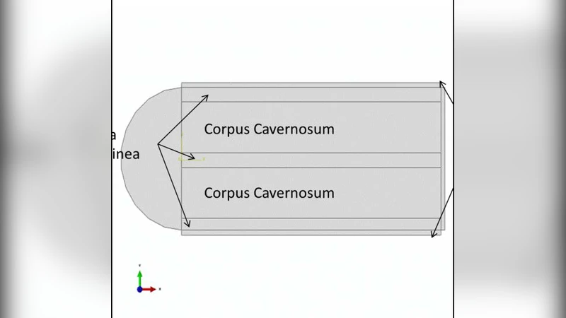
The model geometry replicates an 8 cm long, 4 cm diameter penis in its flaccid state and includes three tissue layers: skin, tunica albuginea, and corpus cavernosum. Material properties were assigned based on literature and previous measurements: skin and tunica albuginea were modeled as transverse‑isotropic elastic solids with shear moduli of 30 kPa and 12 kPa respectively, while the corpus cavernosum was treated as an isotropic porous viscoelastic medium (density = 330 kg/m³, void ratio = 0.7). Linear quadrilateral CPS4R elements of 1 mm × 1 mm were used, with hour‑glass control and reduced integration to mitigate shear locking. Boundary conditions fixed the bottom and right edges; an infinite element layer (CINPE4) surrounded the domain to suppress artificial reflections.
Erectile pressure was applied uniformly to the internal surfaces of the corpora, ranging from 20 mmHg to 100 mmHg in 10 mmHg increments, thereby simulating progressive erection. For each EP level, a 3 mm segment source on the skin surface imposed a vertical displacement of 0.1 mm for 0.1 s, generating harmonic excitations at 100, 150, and 200 Hz. Displacements at eight nodes located 5 mm from the source were recorded. A two‑dimensional Fast Fourier Transform (2D‑FFT) converted space‑time data into k‑space, allowing extraction of wave number (k) and angular frequency (ω). Shear wave speed was calculated as c = ω/k.
Simulation results revealed a clear, non‑linear, positive correlation between EP and SWS across all excitation frequencies. At 20 mmHg, SWS was approximately 2.4 m/s; at 100 mmHg it rose to about 7.7 m/s. Moreover, for a given EP, higher excitation frequencies produced higher SWS, indicating frequency‑dependent viscoelastic behavior of penile tissue. Stress distribution maps showed that the tunica albuginea bears the greatest stress during erection (13–55 kPa), consistent with published biomechanical data, while the corpus cavernosum exhibits comparatively lower stiffness.
The authors discuss the physiological implications: as intracavernosal pressure increases, the smooth‑muscle‑rich corpus expands, collagen fibers become taut, and overall tissue rigidity rises, leading to faster shear wave propagation. The observed non‑linear trend aligns with prior experimental measurements of SWS in human subjects (flaccid ≈ 2.4 m/s, erect ≈ 7.7 m/s). The study also acknowledges several modeling simplifications: (1) treating the corpus cavernosum as a homogeneous isotropic poroviscoelastic material neglects its complex micro‑architecture of collagen, smooth muscle, and endothelium; (2) blood flow and fluid‑structure interaction are omitted, although they are integral to erection dynamics; (3) tissue anisotropy, especially in the tunica albuginea, is not represented. These limitations suggest that future work should incorporate fluid‑structure coupling and anisotropic constitutive models, as well as experimental validation with in‑vivo data.
In conclusion, this work provides the first computational evidence that EP directly influences shear wave speed in penile tissue, supporting the use of UVE as a non‑invasive tool for quantifying erectile function. By enabling indirect measurement of intracavernosal pressure and tissue stiffness, UVE could improve diagnosis, monitor therapeutic response, and potentially serve as an early indicator of cardiovascular risk associated with erectile dysfunction. The authors propose that further refinement of the FEM and clinical studies will solidify UVE’s role in urological practice.
Comments & Academic Discussion
Loading comments...
Leave a Comment