Effects of Important Parameters Variations on Computing Eigenspace-Based Minimum Variance Weights for Ultrasound Tissue Harmonic Imaging
In recent years, the minimum variance (MV) beamforming has been widely studied due to its high resolution and contrast in B-mode Ultrasound imaging (USI). However, the performance of the MV beamformer is degraded at the presence of noise, as a result…
Authors: Mehdi Haji Heidari, Moein Mozaffarzadeh, Rayyan Manwar
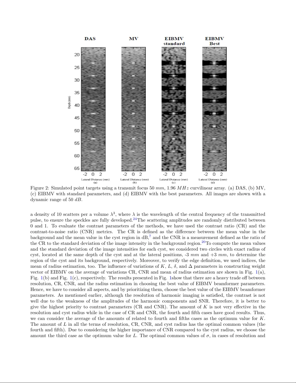
Effects of Imp ortan t P arameters V ariations on Computing Eigenspace-Based Minim um V ariance W eigh ts for Ultrasound Tissue Harmonic Imaging Mehdi Ha ji Heidari a , Mo ein Mozaffarzadeh a,b,* , Ra yyan Man w ar c , and Mohammadreza Nasiria v anaki c a Departmen t of Biomedical Engineering, T arbiat Mo dares Univ ersit y , T ehran, Iran b Researc h Center for Biomedical T ec hnologies and Rob otics, Institute for Adv anced Medical T echnologies, T ehran, Iran c Departmen t of Biomedical Engineering, W a yne State Universit y , Detroit, Michigan, USA ABSTRA CT In recen t years, the minim um v ariance (MV) b eamforming has b een widely studied due to its high resolution and con trast in B-mo de Ultrasound imaging (USI). How ev er, the p erformance of the MV beamformer is degraded at the presence of noise, as a result of the inaccurate co v ariance matrix estimation which leads to a lo w quality image. Second harmonic imaging (SHI) pro vides many adv an tages ov er the con ven tional pulse-ec ho USI, suc h as enhanced axial and lateral resolutions. Ho wev er, the lo w signal-to-noise ratio (SNR) is a ma jor problem in SHI. In this pap er, Eigenspace-based minimum v ariance (EIBMV) beamformer has been emplo yed for second harmonic USI. The Tissue Harmonic Imaging (THI) is achiev ed by Pulse Inv ersion (PI) technique. Using the EIBMV w eights, instead of the MV ones, would lead to reduced sidelobes and improv ed contrast, without compromising the high resolution of the MV b eamformer (even at the presence of a strong noise). In addition, w e ha v e in v estigated the effects of v ariations of the important parameters in computing EIBMV weigh ts, i.e., K , L , and δ , on the resolution and contrast obtained in SHI. The results are ev aluated using n umerical data (using p oin t target and cyst phan toms), and the prop er parameters of EIBMV are indicated for THI. Keyw ords: Tissue harmonic imaging, minimum v ariance, eigenspace-based minim um v ariance, beamforming, con trast resolution. 1. INTR ODUCTION Second Harmonic Imaging (SHI) is a w ell-known ultrasound medical imaging technique that has improv ed im- age quality in many clinical ultrasound applications. 1 The resolution and contrast pro vided b y the harmonic imaging is muc h b etter than the fundamental imaging. 2 , 3 Although the harmonic imaging provides adv antages compared to the fundamental imaging, the amplitudes of the harmonic components are so w eak. Therefore, Tissue Harmonic Imaging (THI) alw ays suffers from a p o or Signal-to-Noise Ratio (SNR). 4 – 6 Beamforming plays a significant role in the quality of the formed images. There are a large n umber of publica- tions in this field of study. 7 – 9 As nov el weigh ting methods, tw o mo difications of the Coherence F actor (CF) hav e b een introduced. 10 , 11 The Minimum V ariance (MV) b eamformer is an adaptiv e w eighting metho d which can b e applied to the medical ultrasound B-mo de images to improv e the resolution. 12 The MV relies on weigh ted summation of received signals in whic h weigh ts are calculated based on the information obtained by the spa- tial prop erties of the recorded signals. There are a num b er of mo difications for MV algorithm. 13 – 18 Adaptiv e b eamforming metho ds used in ultrasound images provides a significant higher resolution. How ev er, the con trast enhancemen t is not inv estigated significan tly . The Eigenspace-Based Minim um V ariance (EIBMV) b eamformer is a tec hnique in whic h not only it can improv e the resolution, but also significan tly enhances the contrast, compared to Delay and Sum (DAS) and MV b eamformers. The weigh t vector of the EIBMV is generated by pro jecting the MV weigh t vector onto a vector subspace constructed from the eigenstructure of the cov ariance *F urther author information: (Send corresp ondence to M. Mozaffarzadeh) Mo ein Mozaffarzadeh: E-mail: mo ein.mfh@modares.ac.ir matrix. 19 In the EIBMV approach, the effective parameters for computing w eights ha ve standard v alues which are generally applied to the MV-based approac hes. 19 In this pap er, the EIBMV metho d has b een applied to SHI in order to o vercome the pow erful noise, resulting in contrast impro vemen t. In addition, we ha ve in v estigated the effects of v ariations of the imp ortan t parameters for computing EIBMV weigh ts on the resolution and contrast in THI, and finally , the appropriate parameters are presen ted. The rest of the pap er is organized as follows. The structure of the EIBMV b eamformer and the pro cedure of c ho osing the appropriate weigh ts vector are explained in the section 2 . The influence of the v ariations of EIBMV parameters along with the optimal v alues for parameters and the corresp onding results are illustrated in the section 3 . Finally , the discussion and conclusion are presen ted in the section 4 . 2. METHODS F or a linear array with M elemen ts, the output of a general b eamformer is defined as follow: z ( k ) = w H ( k ) y ( k ) = M X i =1 w i ∗ ( k ) y i ( k − ∆ i ) , (1) where y ( k ) = [ y 1 ( k − ∆ 1 ) , ..., y M ( k − ∆ M )] is the time dela yed version of arra y observ ation corresp onding to a specific p oin t of the image, ∆ i is the time-dela y applied to c hannel i , w = [ w 1 , ..., w M ] T is the complex v ector of b eamformer w eight, and the sup erscripts ( . ) ∗ and ( . ) H denote the conjugate and conjugate transp ose, resp ectiv ely . In the DAS b eamformer, the weigh ts are predetermined by some windowing functions such as rectangular, Hanning and Hamming windows. In the MV b eamformer, the optim um weigh t vectors are obtained as a function of the spatial cov ariance matrix, R , as follows: w M V = R − 1 a a H R − 1 a , (2) where R ( k )= E { y ( k ) y ( k ) H } is the M × M spatial cov ariance matrix, and a is a vector of all ones with length in accordance with R . In practice, the exact cov ariance matrix, R , is una v ailable. Hence, the co v ariance matrix at time k is estimated by using temp oral a veraging and spatial smo othing techniques, where spatial smo othing is done through division of the array into subarrays to obtain a go o d estimate of the cov ariance matrix. 18 R can b e estimated as follows: ˆ R ( k ) = 1 (2 K +1)( M − L +1) K X n = − K M − L +1 X i =1 y i ( k + n ) y i ( k + n ) H , (3) where L is the subarray length, y i ( k + j ) is the delay ed input vector for the i th subarra y , and K is the temp oral a veraging num b er. T o insure a well-conditioned cov ariance matrix and improv e the b eamformer robustness, the co v ariance matrix is diagonally loaded, ˆ R ( k )= ˆ R ( k )+ ε I , where amoun t of the ε has b een set ∆ times of the pow er in the received signals according to ε = ∆trace { ˆ R ( k ) } , and I is the iden tify matrix. 12 , 20 The MV b eamformer is commonly used in ultrasound imaging in order to impro ve resolution of the images. Ho wev er, the contrast enhancemen t has no salient progress. EIBMV metho d, using the eigenstructure of the cov ariance matrix b esides preserving the desire signal, suppresses the sidelob es and consequently improv es image quality in the terms of con trast and resolution. Hence, EIBMV approach retains the resolution while improv es the contrast, compared to MV and DAS b eamformers. In this technique, the estimated cov ariance matrix is broken to tw o orthogonal spaces i.e., signal subspace and noise plus interference one. The diagonally loaded cov ariance matrix R can b e written in terms of its Eigen decomp osition as: ˆ R = VΛV H , (4) where Λ = diag [ λ 1 , λ 2 , ..., λ L ] so that λ 1 ≥ λ 2 ≥ ... ≥ λ L and V = [ v 1 , v 2 , ..., v L ] in which v i , i = 1 , 2 , ..., L are the orthonormal eigen vectors corresp onding to λ i , i = 1 , 2 , ..., L . T o limit the effects of interferences and noise, the cov ariance matrix can b e generated b y a few num b ers of eigenv ectors. The signal subspace E s only T able 1: Parameters of the Simulation Setup. V alue P arameter 50 M H z Sampling frequency ( f s ) 1.96 M H z Cen ter fundamental frequency ( f 0 ) 3.92 M H z Cen ter fundamental frequency (2 f 0 ) 132 Num b er of transmission elements 66 Num b er of receive elements ( M ) 409 µm Pitc h 20 µm Kerf 1540 m/s sp eed of sound ( C 0 ) 50 mm F ocal depth (transmit fo cus) 1000 k P a Initial pressure 1000 k g /m 3 Medium density 0.5 dB /cm/ M H z atten uation co efficien t 3.5 Nonlinear parameter ( β ) 2 Gamma co efficien t ( γ ) T able 2: Parameters for W eights Calculation. P arameter First Second Third F ourth Fifth K 0 1 3 K stan 1 2 K stan K stan 2 K stan L 1 1 6 M 1 3 M 1 2 M = L stan M ∆ 0 1 100 ∆ stan 1 10 ∆ stan ∆ stan 10∆ stan δ 0 0.1 0.5 0.8 1 con tains the desire signal and significan tly decreases the sidelob es effects. This subspace is constructed from the eigen vectors corresp onding to the largest eigenv alues. E s = [ v 1 , ..., v N um ] (5) where N um is the num b er of the eigenv ectors effectiv ely demonstrate the signal subspace. One metric for determining the N U M could b e defined as the num b er of dominan t eigen v alues, the amoun t of which are larger than δ λ max , where δ is a p ositiv e factor smaller than 1, and λ max is the largest eigenv alue. Finally , the weigh t v ector of the EIBMV is given by pro jecting the MV weigh t vector on to signal subspace. 19 w E I B M V = E s E H s w M V (6) 3. RESUL TS 3.1 Data Acquisition In b oth MV and EIBMV adaptive b eamformers, the effective parameters for weigh ts calculation hav e standard v alues; these parameters are: (1) the n umber of samples for the temp oral av eraging of the cov ariance matrix ( K ), (2) the num b er of subarra y elements for computing the cov ariance matrix ( L ), (3) the diagonal loading factor (∆) , and (4) In EIBMV approach the δ parameter, which their standard v alues are the length of the transmission pulse ( K stan ), half of the num b er of arra y elemen ts ( L = M / 2), ( K stan = 1 / 100 L ), and δ stan = 0 . 5, resp ectiv ely. 19 In this study , we aim to inv estigate the influence of all the four effective parameters of EIBMV w eights calculation on the resolution and contrast, for the SHI, in order to c ho ose their b est v alue. In this section, we hav e simulated t wo phan toms, a phantom of wire targets lo cated at the depth of 22.5-62.5 mm , separated b y 5 mm at eac h depth, and a phan tom including 5 cysts with an equal diameter of 5 mm , lo cated at the depth of 22.5-62.5 mm , separated by 10 mm . The phan toms are used in order to inv estigate the p erformance of the EIBMV b eamformer in the terms of the lateral resolution and contrast, along with changes of K , L , ∆ and σ . The simulations hav e b een carried out using CREANUIS, whic h is a non-linear radio frequency (RF) Figure 1: The patterns obtained from the a v erage of (a) CR, (b) CNR, and (c) estimate d cyst radius (for the sim ulated c yst phan tom along the depths of 22.5-62.5 mm ), and (d) the a v erage of lateral FWHM (for 9 p oin ts along the depths of 22.5-62.5 mm ) for v ariations of the K , L , δ , and ∆ par a meters in the w eigh t v ector construction, using a transmit fo c u s of 50 mm , 1.96 M H z curvilinear arra y . The amoun ts of K , L , δ , and ∆ parameter considered for computing w e igh ts are listed at T able 2 . ultrasound image sim ulator. 21 , 22 In the all sim ulations, a 132-elemen t curvilin e ar arra y transdu c er w ere used. In transmission, all eleme n ts are participated while in reception only the nearest 66 elemen ts of the transducer whic h w ere placed in fron t of the sc an line w ere used. A t w o-cycle Gaussian w eigh ted S in usoidal signal w as used in transmission, and the transmit fo cus w as set at 50 mm depth. Other parameters for sim u lations are listed in T able 1 . In all the sim ulations, to separate the harmonic fr o m fundamen tal frequencies, w e ha v e used PI tec hnique whi c h is a w ell-kno wn tec hnique. 23 Before separating the harmonic from fundamen tal b y the PI metho d, white Gaussian noise with a SNR of 60 dB w as added to the receiv ed signal. In the MV and EIBMV b eamformers, the am ou n ts of parameters considered for computing w eigh ts are listed at T able 2 . 3.2 Sim ulated P oin t T argets Here, w e ai m to explore th e influence of v ariations of the effectiv e parameters of the EIBMV on the resolution of the output image. W e ha v e used the F ull Width at Half Maxim um (FWHM) index to ev aluate the mainlob e width of the images for eac h p oin t. Fig. 1 (d) sho ws the influence of v ariations of K , L , ∆ and δ on the a v erage of the mainlob e width (in the term of FWHM), for all the target p oin ts. It is notable that a less mainlob e width for a p oin t in a sp ecific depth indicates a high e r resolution. 3.3 Sim ulated C ys t T argets T o in v estigate the i nfluence of the v ariations of the eff ectiv e parameters in EIBMV, in the term of con trast, a phan tom of c ysts w ere used. In sim ulating the cyst phan tom, the sp ec kle distribution is sim ulated rand om l y with Figure 2: Sim ulated p oin t targets using a transmit fo c u s 50 mm , 1.96 M H z curvilinear arra y . (a) D AS, (b) MV, (c) EIBMV with stand ard parameters, and (d) EIBMV with th e b es t parameters. All images are sho wn with a dynamic range of 50 dB . a densit y of 10 scatters p er a v olu m e λ 3 , where λ is the w a v elength of the cen tral fr e qu e n c y of the transmitted pulse, to ensure the sp ec kles are fully dev elop ed. 24 The scattering amplitudes are randomly distributed b et w een 0 and 1. T o ev aluate the con trast parameters of the metho ds, w e ha v e used the con trast ratio (CR) and the con trast-to-noise ratio (CNR) metrics. The CR is defin e d as the diffe r e n c e b et w een th e mean v alue in the bac kground and the mean v alue in th e cyst region in dB, 7 and the CNR is a me asuremen t defined as the rat io of the CR to the standard deviation of the image in tensit y in the bac kground region. 20 T o compute the me an v alues and the standard deviation of the image in tensities for eac h cyst, w e considered t w o circles with exact radius of cyst, lo cated at the same depth of the cyst and at the lateral p ositions, -3 mm and +3 mm , to determine the region of the cyst and its bac kground, resp ectiv ely . Moreo v e r, to v erify the edge definition, w e used indices, the mean of radius estimation, to o. The influence of v ariations of K , L , δ , and ∆ parameters in constructing w eigh t v ector of EIBMV on the a v erage of v ariations CR, CNR and mean of radius estimation are sho wn in Fig. 1 (a), Fig. 1 (b) and Fig. 1 (c), resp ectiv ely . The res u lts presen ted in Fig. 1 sho w that there are a hea vy trade off b et w een resolution, CR, CNR, and the radius estimation in c ho osing the b est v alue of EIBMV b eamformer parameters. Hence, w e ha v e to consider all asp ects, and b y prioritizing them, c ho ose the b est v alue of the EIBMV b eamformer parameters. As men tioned earlier, although the resolution of harmonic imaging is satisfied, the con trast is not w ell due to the w e akn e ss of the amplitu des of th e h armonic c omp onen t s and SNR. Therefore, it is b etter to giv e the highest priorit y to con trast parameters (CR and CNR). The amoun t of K is not v ery eff ectiv e in the resolution and cyst radius while in the case of CR and CNR, the fourth and fifth cases ha v e go o d results. Th us, w e can consider the a v erage of the amoun ts of related to fourth and fifths cases as the optim um v alue for K . The amoun t of L in all the terms of resolution, CR, CNR, and cys t radius has the optimal common v alues (the fourth and fifth). Due to considering the higher im p ortance of CNR com p ared to the cyst radius, w e c ho ose the amoun t the third case as the optim um v alue for L . The optimal common v alues of σ , in cases of r e soluti on and Figure 3: Boundaries of sim ulated cyst phan tom using a transmit fo cus 50 mm , 1.96 M H z curvilinear arra y . (a) D AS, (b) MV, (c) EIBMV with standard parameters, and (d) EIBMV with b est parameters. The panels from top to b ottom are cyst lo cate d at depths 22.5 mm , 32.5 mm , 42.5 mm , 52.5 mm , and 62.5 mm , resp ectiv ely . All the images are sho wn with a dynamic range of 50 dB . cyst radius are the v alues of the fourth an d fifth cases, while for CR and CNR, the v alues are selected based on the second and third cas es. Due to considerin g a higher imp ortance for CR an d CNR, compared to resolution and cyst radius parameter, w e c ho ose the amoun t related to third case as the optim um v alue for δ , whic h is the optimal common v alue among all the resolution and con trast paramete r s . The ∆ parameter do es not ha v e significan t effect on the resolution and con tr as t par am eters. So, w e totally ignore analysis of this parameter; and set its optimal v alue as its standard v alue. Results demonstrate that the b est v alues of parame ters K , L , ∆ and δ for the EIBMV b eamformer are K best = 1 . 5 K stan , L best = 2 / 3 L stan = 1 / 3 M , ∆ best = ∆ stan and δ best = δ stan , Figure 4: The patterns of (a) Lateral FWHM for 9 p oin ts along depths 22.5-62.5 mm , and the patterns of (b) CR, (c) CNR, an d (d) estimated cyst radius, from sim ulated cyst phan tom alon g depths 22.5-62.5 mm , resp ectiv ely , for (a) D AS, (b) MV, (c) EIBMV with standard parameters, and (d) EIBMV with b est parameters, u s i ng a transmit fo cus 50 mm , 1.96 M H z curvilinear arra y . resp ectiv ely . 4. DISCUSSION AND CONCLUSION Harmonic imaging has go o d axial and lateral resolutions. Ho w ev er, the lo w SNR is a ma jor prob le m in v olv ed with this imaging m etho d. In this pap er, EIBMV b eamformer is ap plied to SHI. Using EIBMV w eigh ts instead of the MV ones, leads to reduced sidelob es and impro v e d con trast, without compromising the high resolution of the MV b eamforme r , ev e n at the presence of strong noise. Moreo v er, here w e in v estigated the effects of v ariations of the imp ortan t parameters in computing EIBMV w eigh ts, i.e., K , L , ∆ and δ , on the resolution and con trast while THI is p erformed. T o this end, the effects of eac h parameter is separately explored while other parameters w ere fixed (ha ving their standard v alues). The results, using p oin t target and cyst p han tom, are ev aluated on n umerical data, and the prop er parameters of EIBMV are indicated for THI. Results demonstrate that the b est v alues of K , L , ∆ and δ parameters for EIBMV b eamformer are K best = 1 . 5 K stan , L best = 2 / 3 L stan = 1 / 3 M , ∆ best = ∆ stan and δ best = δ stan , resp ectiv ely . Fig. 2 and Fig. 3 sho w the images obtained from th e men tioned phan toms in the s ection 2 , p oin t targets and c y s t phan tom, resp ectiv ely , using differen t b eamformers (D AS, MV, EIBMV with standard parameters, and EIBMV with the b est parameters), displ a y ed o v er a 50 dB dynamic range. In Fig. 3 the b ound aries of the sim ulated cyst phan toms are displ a y ed, at all depth, o v er a 30 dB dynamic range. The red circles and the green curv es plotted in Figs. 3 represen t the true and estimated b order of the cysts, r e sp ectiv ely . The FWHM v ariation along the depths for all p oin ts of Fig. 2 , and also the v ariations of relativ e CR, CNR, m ean of radius estimation, along the depths for all cyst targets of Fig. 3 , are sho wn in Fig. 4 , resp ectiv ely . As the results sho w, although EIBMV metho d, where the prop osed parameter are used instead of the standard parameter, decreases the abilit y to impro v e the resolution and cyst radius, it impro ves CR and CNR significan tly . In other w ords, the EIBMV beamformer could obtain a better con trast by sacrificing the resolution, whic h w ould b e suitable for harmonic imaging. The presented results show that, in av erage, EIBMV metho d when the prop osed parameter are used provides CR enhancemen t of 10.6 dB , 9.2 dB , and 0.4 dB , and CNR impro vemen t of ab out 78%, 62%, and 32% in comparison with DAS, MV and EIBMV with standard parameter, resp ectiv ely . It would b e more efficient to implement the EIBMV technique with the MV metho ds having a low computational complexities 25 – 29 instead of using the conv entional MV. Therefore, a practical metho d can b e ac hieved. This is a topic for our future work. REFERENCES [1 ] Li, Y. and Zagzebski, J. A., “Computer mo del for harmonic ultrasound imaging,” IEEE tr ansactions on ultr asonics, ferr o ele ctrics, and fr e quency c ontr ol 47 (4), 1000–1013 (2000). [2 ] Shen, C.-C. and Li, P .-C., “Harmonic leak age and image quality degradation in tissue harmonic imaging,” IEEE tr ansactions on ultr asonics, ferr o ele ctrics, and fr e quency c ontr ol 48 (3), 728–736 (2001). [3 ] Bouak az, A. and de Jong, N., “Native tissue imaging at sup erharmonic frequencies,” IEEE tr ansactions on ultr asonics, ferr o ele ctrics, and fr e quency c ontr ol 50 (5), 496–506 (2003). [4 ] Du, Y., Rasmussen, J., Jensen, H., and Jensen, J. A., “Second harmonic imaging using synthetic ap erture sequen tial beamforming,” in [ Ultr asonics Symp osium (IUS), 2011 IEEE International ], 2261–2264, IEEE (2011). [5 ] Christopher, T., “Finite amplitude distortion-based inhomogeneous pulse echo ultrasonic imaging,” IEEE tr ansactions on ultr asonics, ferr o ele ctrics, and fr e quency c ontr ol 44 (1), 125–139 (1997). [6 ] Mardi, Z. and Mahlo o jifar, A., “Tissue second harmonic ultrasound imaging using huffman sequence,” in [ Biome dic al Engine ering and 2016 1st International Ir anian Confer enc e on Biome dic al Engine ering (ICBME), 2016 23r d Ir anian Confer enc e on ], 182–186, IEEE (2016). [7 ] Mozaffarzadeh, M., Mahlo o jifar, A., Oro o ji, M., Adabi, S., and Nasiriav anaki, M., “Double-stage delay m ul- tiply and sum b eamforming algorithm: Application to linear-arra y photoacoustic imaging,” IEEE T r ansac- tions on Biome dic al Engine ering 65 (1), 31–42 (2018). [8 ] Mozaffarzadeh, M., Mahlo o jifar, A., and Oro o ji, M., “Image enhancement and noise reduction using mo di- fied delay-m ultiply-and-sum b eamformer: Application to medical photoacoustic imaging,” in [ Ir anian Con- fer enc e on Ele ctric al Engine ering (ICEE) 2017 ], 65–69, IEEE (2017). [9 ] Mozaffarzadeh, M., Sadeghi, M., Mahlo o jifar, A., and Oro o ji, M., “Double-stage delay multiply and sum b eamforming algorithm applied to ultrasound medical imaging,” Ultr asound in Me dicine and Biolo gy 44 (3), 677 – 686 (2018). [10 ] Mozaffarzadeh, M., Y an, Y., Mehrmohammadi, M., and Makkiabadi, B., “Enhanced linear-array photoa- coustic b eamforming using mo dified coherence factor,” Journal of Biome dic al Optics 23 (2), 026005 (2018). [11 ] Mozaffarzadeh, M., Mehrmohammadi, M., and Makkiabadi, B., “Image improv ement in linear-array photoacoustic imaging using high resolution c oh e rence factor weigh ting technique,” arXiv pr eprint arXiv:1710.02751 (2017). [12 ] N¨ asholm, S. P ., Austeng, A., Jensen, A. C., Nilsen, C.-I. C., and Holm, S., “Cap on b eamforming applied to second-harmonic ultrasound exp erimental data,” in [ Ultr asonics Symp osium (IUS), 2011 IEEE Interna- tional ], 2217–2220, IEEE (2011). [13 ] Mozaffarzadeh, M., Mahlo o jifar, A., and Oro o ji, M., “Medical photoacoustic b eamforming using minimum v ariance-based dela y multiply and sum,” in [ Digital Optic al T e chnolo gies 2017 ], 10335 , 1033522, Interna- tional So ciet y for Optics and Photonics (2017). [14 ] Mozaffarzadeh, M., Av anji, S. A. O. I., Mahlo o jifar, A., and Oro o ji, M., “Photoacoustic imaging using com bination of eigenspace-based minimum v ariance and delay-m ultiply-and-sum b eamformers: Simulation study ,” arXiv pr eprint arXiv:1709.06523 (2017). [15 ] Paridar, R., Mozaffarzadeh, M., Mehrmohammadi, M., and Oro o ji, M., “Photoacoustic image formation based on sparse regularization of minimum v ariance b eamformer,” arXiv pr eprint arXiv:1802.03724 (2018). [16 ] Paridar, R., Mozaffarzadeh, M., Nasiriav anaki, M., and Oro o ji, M., “Double minimum v ariance b eamform- ing metho d to enhance photoacoustic imaging,” arXiv pr eprint arXiv:1802.03720 (2018). [17 ] Mozaffarzadeh, M., Mahlo o jifar, A., Oroo ji, M., Kratkiewicz, K., Adabi, S., and Nasiria v anaki, M., “Linear- arra y photoacoustic imaging using minimum v ariance-based delay multiply and sum adaptive b eamforming algorithm,” Journal of Biome dic al Optics 23 (2), 026002 (2018). [18 ] Mozaffarzadeh, M., Mahlo o jifar, A., Nasiria v anaki, M., and Oro o ji, M., “Eigenspace-based minim um v ari- ance adaptive b eamformer combined with delay m ultiply and sum: Exp erimen tal study ,” arXiv pr eprint arXiv:1710.01767 (2017). [19 ] Asl, B. M. and Mahlo o jifar, A., “Eigenspace-based minimum v ariance b eamforming applied to medical ultrasound imaging,” IEEE tr ansactions on ultr asonics, ferr o ele ctrics, and fr e quency c ontr ol 57 (11) (2010). [20 ] Asl, B. M. and Mahlo o jifar, A., “Minim um v ariance beamforming com bined with adaptive coherence w eight- ing applied to medical ultrasound imaging,” IEEE tr ansactions on ultr asonics, ferr o ele ctrics, and fr e quency c ontr ol 56 (9) (2009). [21 ] V arra y , F., Ramalli, A., Cachard, C., T ortoli, P ., and Basset, O., “F undamental and second-harmonic ultrasound field computation of inhomogeneous nonlinear medium with a generalized angular sp ectrum metho d,” IEEE tr ansactions on ultr asonics, ferr o ele ctrics, and fr e quency c ontr ol 58 (7) (2011). [22 ] V arra y , F., Basset, O., T ortoli, P ., and Cachard, C., “Creanuis: a non-linear radiofrequency ultrasound image simulator,” Ultr asound in me dicine & biolo gy 39 (10), 1915–1924 (2013). [23 ] Ma, Q., Ma, Y., Gong, X., and Zhang, D., “Impro vemen t of tissue harmonic imaging using the pulse- in version technique,” Ultr asound in me dicine & biolo gy 31 (7), 889–894 (2005). [24 ] Holfort, I. K., Gran, F., and Jensen, J. A., “Broadband minim um v ariance b eamforming for ultrasound imaging,” IEEE tr ansactions on ultr asonics, ferr o ele ctrics, and fr e quency c ontr ol 56 (2), 314–325 (2009). [25 ] Deylami, A. M. and Asl, B. M., “A fast and robust b eamspace adaptive beamformer for medical ultrasound imaging,” IEEE T r ansactions on Ultr asonics, F err o ele ctrics, and F r e quency Contr ol (2017). [26 ] Nguyen, M. M., Shin, J., and Y en, J., “Harmonic imaging with fresnel beamforming in the presence of phase ab erration,” Ultr asound in me dicine & biolo gy 40 (10), 2488–2498 (2014). [27 ] Asl, B. M. and Mahlo o jifar, A., “A lo w-complexity adaptiv e b eamformer for ultrasound imaging using structured cov ariance matrix,” IEEE tr ansactions on ultr asonics, ferr o ele ctrics, and fr e quency c ontr ol 59 (4) (2012). [28 ] Moghimirad, E., Hoy os, C. A. V., Mahlo o jifar, A., Asl, B. M., and Jensen, J. A., “Syn thetic ap erture ultrasound fourier b eamformation using virtual sources,” IEEE tr ansactions on ultr asonics, ferr o ele ctrics, and fr e quency c ontr ol 63 (12), 2018–2030 (2016). [29 ] Deylami, A. M. and Asl, B. M., “Lo w complex subspace minimum v ariance b eamformer for medical ultra- sound imaging,” Ultr asonics 66 , 43–53 (2016).
Original Paper
Loading high-quality paper...
Comments & Academic Discussion
Loading comments...
Leave a Comment