Title: Shared High Value Research Resources: The CamCAN Human Lifespan Neuroimaging Dataset Processed on the Open Science Grid
ArXiv ID: 1710.05246
Date: 2017-12-11
Authors: Researchers from original ArXiv paper
📝 Abstract
The CamCAN Lifespan Neuroimaging Dataset, Cambridge (UK) Centre for Ageing and Neuroscience, was acquired and processed beginning in December, 2016. The referee consensus solver deployed to the Open Science Grid was used for this task. The dataset includes demographic and screening measures, a high-resolution MRI scan of the brain, and whole-head magnetoencephalographic (MEG) recordings during eyes closed rest (560 sec), a simple task (540 sec), and passive listening/viewing (140 sec). The data were collected from 619 neurologically normal individuals, ages 18-87. The processed results from the resting recordings are completed and available online. These constitute 1.7 TBytes of data including the location within the brain (1 mm resolution), time stamp (1 msec resolution), and 80 msec time course for each of 3.7 billion validated neuroelectric events, i.e. mean 6.1 million events for each of the 619 participants. The referee consensus solver provides high yield (mean 11,000 neuroelectric currents/sec; standard deviation (sd): 3500/sec) high confidence (p < 10-12 for each identified current) measures of the neuroelectric currents whose magnetic fields are detected in the MEG recordings. We describe the solver, the implementation of the solver deployed on the Open Science Grid, the workflow management system, the opportunistic use of high performance computing (HPC) resources to add computing capacity to the Open Science Grid reserved for this project, and our initial findings from the recently completed processing of the resting recordings. This required 14 million core hours, i.e. 40 core hours per second of data.
💡 Deep Analysis
Deep Dive into Shared High Value Research Resources: The CamCAN Human Lifespan Neuroimaging Dataset Processed on the Open Science Grid.
The CamCAN Lifespan Neuroimaging Dataset, Cambridge (UK) Centre for Ageing and Neuroscience, was acquired and processed beginning in December, 2016. The referee consensus solver deployed to the Open Science Grid was used for this task. The dataset includes demographic and screening measures, a high-resolution MRI scan of the brain, and whole-head magnetoencephalographic (MEG) recordings during eyes closed rest (560 sec), a simple task (540 sec), and passive listening/viewing (140 sec). The data were collected from 619 neurologically normal individuals, ages 18-87. The processed results from the resting recordings are completed and available online. These constitute 1.7 TBytes of data including the location within the brain (1 mm resolution), time stamp (1 msec resolution), and 80 msec time course for each of 3.7 billion validated neuroelectric events, i.e. mean 6.1 million events for each of the 619 participants. The referee consensus solver provides high yield (mean 11,000 neuroelec
Don Krieger Neurological Surgery
University of Pittsburgh
Pittsburgh, PA, USA
kriegerd@upmc.edu
Paul Shepard Physics and Astronomy University of Pittsburgh
Pittsburgh, PA, USA
shepard@pitt.edu Ben Zusman
Neurological Surgery University of Pittsburgh
Pittsburgh, PA, USA zusmanbe@upmc.edu Anirban Jana
Pittsburgh Supercomputing
Center
Pittsburgh, Pa
anirbanjana@cmu.edu
David Okonkwo
Neurological Surgery
University of Pittsburgh
Pittsburgh, PA, USA
okonkwodo@upmc.edu
Abstract—The CamCAN Lifespan Neuroimaging Dataset
[1,2], Cambridge (UK) Centre for Ageing and Neuroscience, was
acquired and processed beginning in December, 2016. The referee
consensus solver deployed to the Open Science Grid [3,4] was used
for this task. The dataset includes demographic and screening
measures, a high-resolution MRI scan of the brain, and whole-
head magnetoencephalographic (MEG) recordings during eyes
closed rest (560 sec), a simple task (540 sec), and passive
listening/viewing (140 sec). The data were collected from 619
neurologically normal individuals, ages 18-87. The processed
results from the resting recordings are completed and available
for download at http://stash.osgconnect.net/+krieger/
. These
constitute ≈1.7 TBytes of data including the location within the
brain (1 mm resolution), time stamp (1 msec resolution), and 80
msec time course for each of 3.7 billion validated neuroelectric
events, i.e. mean 6.1 million events for each of the 619 participants.
The referee consensus solver provides high yield (mean 11,000
neuroelectric currents/sec; standard deviation (sd): 3500/sec) high
confidence (p < 10-12 for each identified current) measures of the
neuroelectric currents whose magnetic fields are detected in the
MEG recordings. We describe the solver, the implementation of
the solver deployed on the Open Science Grid, the workflow
management system, the opportunistic use of high performance
computing (HPC) resources to add computing capacity to the
Open Science Grid reserved for this project, and our initial
findings from the recently completed processing of the resting
recordings. This required ≈14 million core hours, i.e. ≈40 core
hours per second of data.
Keywords—magnetoencephalography, MEG, referee consensus,
opportunistic computing, shared data, concussion, TBI
I. INTRODUCTION
It has been the informed expectation for a century that the
keys to understanding the human brain will be found in
measuring and understanding the electrical activity of neurons.
Today, clinical neurophysiologists routinely measure single
neurons to aide implantation of therapeutic devices deep in the
brain [5]. Epileptologists use arrays of implanted “stereo EEG”
electrodes and the population recordings obtained from them to
diagnose and guide the treatment of intractable seizure disorders
[6].
It is population activity which is thought to be the basis for
brain function. Stereo EEG and comparable invasive methods
produce voltage recordings with resolution of a few millimeters
at best from up to a few hundred recording sites. Because the
electric field interacts strongly with the conducting tissue in the
brain, these measures are difficult to localize if there is much
tissue between the field source and the electrodes. This problem
is particularly pronounced when the recordings are made
noninvasively from electrodes placed on the scalp.
Magnetoencephalography (MEG) provides an alternative
noninvasive measurement approach with several advantages
over scalp and even implanted EEG recordings. A typical MEG
scanner is shown in Figure 1. The magnetic fields produced by
minute electric currents within the brain are measured at the
MEG sensor array with high fidelity. Unlike electric fields,
magnetic fields do not interact with brain tissue; they pass
through it as if it weren’t there. So in principle, the current which
is the source of such a field is more readily localized. If the
contribution to the MEG measurements of a single neuroelectric
current source can be identified, the corresponding current can
be accurately estimated using the Biot-Savart law [7].
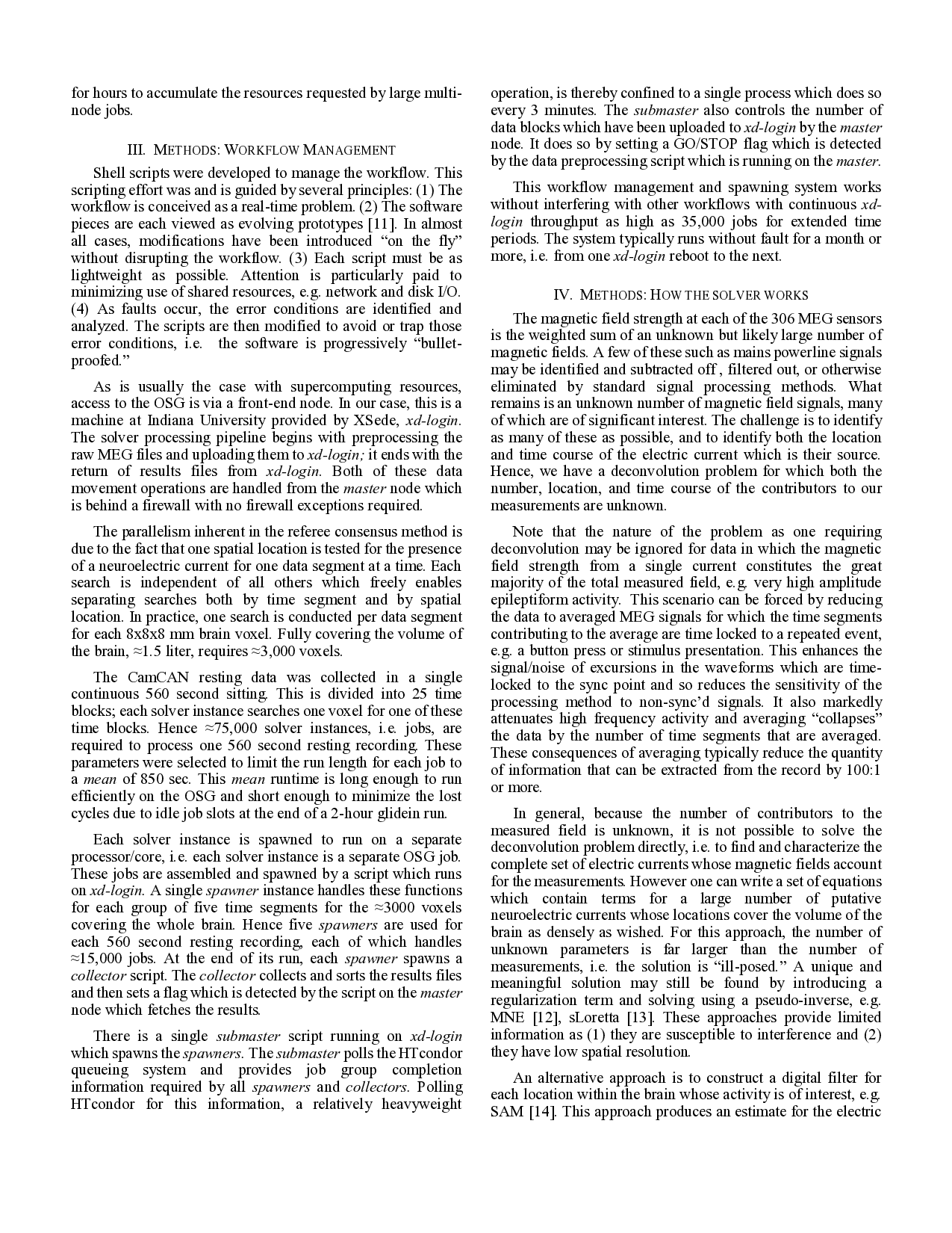
Fig 1. 306 channel whole head VectorView MEG scanner (Elekta, Inc.,
Stockholm, Sweden). The sensor array is composed of 102 planar “chips,”
each with 3 superconducting magnetic field sensors: one magnetometer, two
figure-eight gradiometers at right angles to each other, and a
superconducting quantum interference device (SQUID) coupled to each.
The SQUID’s are used to couple the minute currents induced by magnetic
flux in the sensors to the room temperature electronics.
The measurements at the MEG sensor array are due to an
unknown and almost certainly large number of neuroelectric
currents.