Absence of Typical Haversian System from the Compact Bone of Some Reptile and Bird Species
Background and Objective: Mammalian compact bone is composed mostly of Haversian system. Although there are many studies describing the typical Haversian system in mammals, there are few studies conducted on bones from non-mammalian species. The objective of the current study was to investigate the existence of the typical Haversian system in compact bones from reptiles and birds. Materials and Methods: Femora were collected from geckos, Nile monitors, sparrows, ducks and geese. Samples were then, fixed in 10% paraformaldehyde and embedded in paraffin. Paraffin sections were stained with hematoxylin and eosin or Von Kossa and then examined using light microscopy. Results: The current study showed different histological structures of compact bones from studied species and an absence of the typical Haversian system seen in mammals. The compact bone of geckos was mostly avascular, while Nile monitor bone was highly vascular. Sparrow bone showed one side vascular and the other side avascular. The vascularity was represented by primary vascular canals surrounded by a few irregularly arranged osteocytes inside their lacunae forming primary osteons. Bone tissue from ducks and geese were similar and were composed mostly of dense Haversian tissue, while some areas had primary osteons. Conclusion: The compact bone microstructure in some reptile and bird species lack the typical Haversian system described in mammals. This will be of a great importance in developing a better understanding of long bone anatomy and physiology in different species.
💡 Research Summary
The present study set out to determine whether the classic Haversian system, which dominates the compact bone of mammals, is also a universal feature of the long bones of non‑mammalian vertebrates. To this end, femora from five species representing two reptilian groups (the gecko Hemidactylus sp. and the Nile monitor Varanus niloticus) and three avian groups (the house sparrow Passer domesticus, the domestic duck Anas platyrhynchos, and the domestic goose Anser anser) were harvested, fixed in 10 % paraformaldehyde, embedded in paraffin, sectioned at 5 µm, and stained with hematoxylin‑eosin (H&E) and Von Kossa. Light‑microscopic analysis revealed striking inter‑taxonomic differences in vascularization, osteon architecture, and mineral deposition, all of which contrast sharply with the tightly organized secondary osteons that characterize mammalian compact bone.
In the gecko, compact bone was essentially avascular. No distinct vascular canals or secondary osteons were observed; instead, the matrix consisted of lamellar bone with sparsely distributed lacunae containing osteocytes. This paucity of vascular channels aligns with the low metabolic rate and small body size typical of many small lizards, suggesting that a minimal blood supply is sufficient for bone maintenance in this species.
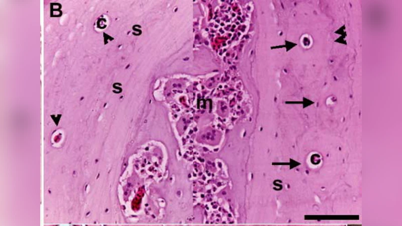
By contrast, the Nile monitor displayed a highly vascularized compact bone. Numerous primary vascular canals traversed the cortex, each surrounded by irregularly arranged osteocytes within lacunae, forming what the authors term “primary osteons.” These structures lacked the concentric lamellae and cement lines that define true secondary osteons, indicating that the monitor’s bone relies on a more primitive, primary remodeling system, perhaps to accommodate its larger size and higher metabolic demands.
The sparrow presented a mixed pattern: one cortical surface was richly supplied with vascular canals, while the opposite surface was almost avascular. This asymmetry may reflect functional adaptations related to flight mechanics, where differential loading could drive localized remodeling. The avascular side still contained scattered primary osteons, suggesting that even in regions with limited blood flow, bone turnover proceeds via primary vascular channels.
Both the duck and the goose exhibited compact bone that the authors describe as “dense Haversian tissue.” However, this tissue differed from mammalian Haversian bone in several respects. The osteons were larger, the lamellar organization was less regular, and many osteocytes were arranged irregularly within their lacunae. Moreover, primary osteons were interspersed throughout the cortex, indicating that secondary remodeling is not the dominant process. Von Kossa staining demonstrated extensive calcium deposition, consistent with the high bone mineral density required for the mechanical stresses of flight and, in the case of waterfowl, buoyancy control.
Collectively, these findings demonstrate that the classic Haversian system—characterized by concentric lamellae, cement lines, and well‑defined secondary osteons—is largely absent in the compact bone of the examined reptiles and birds. Instead, these taxa rely on a mosaic of primary vascular canals, irregular osteon arrangements, and variable degrees of mineralization. The study underscores the importance of recognizing that bone microarchitecture is highly plastic and closely tied to phylogeny, ecology, and functional demands.
Methodologically, the work validates traditional histological techniques (fixation, paraffin embedding, H&E and Von Kossa staining) as effective tools for comparative bone research, while also highlighting the need for complementary approaches such as scanning electron microscopy, micro‑computed tomography, and nano‑indentation to quantify the mechanical implications of the observed microstructural differences.
In conclusion, the absence of a fully developed Haversian system in the compact bone of the selected reptile and bird species expands our understanding of vertebrate skeletal biology. It suggests that the mammalian model of secondary osteonal remodeling is not a universal vertebrate blueprint but rather a lineage‑specific adaptation. This insight has ramifications for evolutionary biology, comparative anatomy, and the selection of appropriate animal models in orthopedic and biomaterials research.
Comments & Academic Discussion
Loading comments...
Leave a Comment