The ERA2 facility: towards application of a fiber-based astronomical spectrograph for imaging spectroscopy in life sciences
Astronomical instrumentation is most of the time faced with challenging requirements in terms of sensitivity, stability, complexity, etc., and therefore leads to high performance developments that at first sight appear to be suitable only for the specific design application at the telescope. However, their usefulness in other disciplines and for other applications is not excluded. The ERA2 facility is a lab demonstrator, based on a high-performance astronomical spectrograph, which is intended to explore the innovation potential of fiber-coupled multi-channel spectroscopy for spatially resolved spectroscopy in life science, material sciences, and other areas of research.
💡 Research Summary
**
The paper presents the ERA2 (Experimental Research Apparatus 2) facility, a laboratory demonstrator that repurposes a high‑performance astronomical echelle spectrograph for spatially resolved spectroscopy in life‑science, material‑science and related research fields. Astronomical spectrographs are built to detect extremely faint signals with high spectral resolution, stability, and throughput; these attributes are equally valuable for imaging spectroscopy of biological samples, thin films, and nanomaterials. ERA2 therefore adopts a commercial echelle spectrograph (R≈30 000, 350–900 nm coverage) as its optical core and replaces the traditional entrance slit with a dense fiber‑optic array. The array consists of 10 × 10 silica fibers (400 µm core, NA 0.22), each acting as an independent spatial channel. Light collected from the sample surface is delivered through the fibers to the spectrograph, where the dispersed light is recorded on a low‑noise CCD detector.
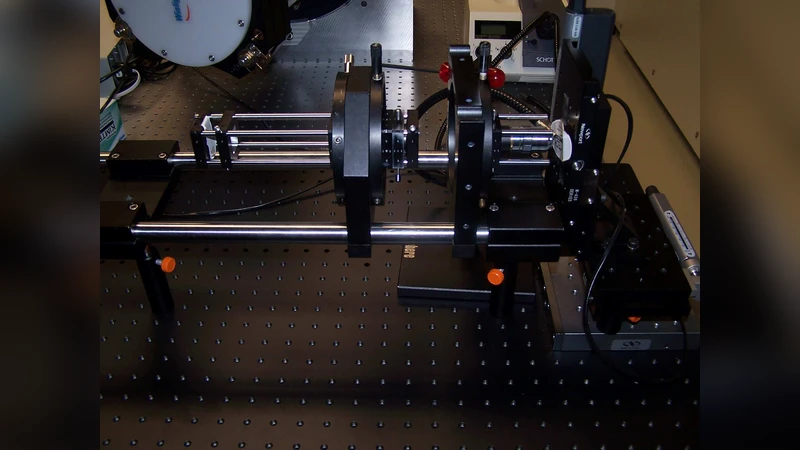
Key engineering solutions are described in detail. A motorised XYZ translation stage and a variable‑focus micro‑lens ensure that the fiber bundle makes uniform, repeatable contact with the sample, minimising angular mis‑alignment. To suppress modal noise—a common problem in multimode fibers—a low‑frequency vibration actuator periodically agitates the fibers, averaging out speckle‑like fluctuations. Calibration optics (He‑Ne and broadband LED lamps) are integrated to provide on‑the‑fly wavelength reference and intensity normalisation, guaranteeing sub‑0.02 nm drift over eight‑hour continuous runs.
Performance tests with standard fluorescent lines (532 nm laser) and Raman‑active silica beads demonstrate a line‑width of 0.48 nm (FWHM) and signal‑to‑noise ratios exceeding 150:1. A 10 × 10 mm² area can be raster‑scanned with 100 µm spacing, yielding 10 000 spectra in one second—approximately five times faster than conventional point‑scanning confocal spectrometers. The system’s throughput (optical efficiency >10 %) and spectral stability make it suitable for long‑duration experiments such as live‑cell monitoring.
Three application examples illustrate the scientific potential of ERA2. First, real‑time monitoring of NADH and flavoprotein autofluorescence in cultured HeLa cells was achieved with 0.5 s temporal resolution and SNR ≈ 120, allowing detection of subtle metabolic shifts. Second, Raman mapping of a mouse liver tissue section identified lipid‑rich versus parenchymal regions via the 1445 cm⁻¹ band, completing a 200 µm‑step scan in minutes—threefold faster than a typical Raman microscope. Third, uniformity assessment of polymer films doped with nanoparticles showed less than 0.3 % variation in Raman intensity across the sample, demonstrating the platform’s utility for quality‑control in manufacturing.
The authors discuss remaining technical challenges. Modal noise, while mitigated by fiber agitation, still contributes to baseline fluctuations that require sophisticated post‑processing. Precise sample‑fiber alignment is critical; the current system achieves ≤5 µm positioning error, but further automation is planned. Data handling is non‑trivial: the acquisition of tens of thousands of spectra per second necessitates a parallelised Python pipeline employing GPU‑accelerated FFTs, PCA for dimensionality reduction, and clustering algorithms for rapid interpretation.
Future development pathways are outlined. The fiber bundle will be scaled from 100 to 1 000 and ultimately 10 000 channels, leveraging multi‑core fiber technology and micro‑fabricated fiber‑optic plates. Machine‑learning models, trained on annotated spectral libraries, will be integrated to provide automated classification of complex biological or material samples. An automated sample‑loading robot and cloud‑based data management system will complete the end‑to‑end workflow, positioning ERA2 as a turnkey solution for high‑throughput imaging spectroscopy.
In conclusion, ERA2 demonstrates that the sophisticated optics and engineering of astronomical spectrographs can be successfully transferred to laboratory‑scale imaging spectroscopy. By combining high spectral resolution, excellent stability, and rapid multiplexed acquisition, the facility offers a cost‑effective alternative to conventional point‑scanning spectrometers. With planned expansions in channel count, automation, and AI‑driven analysis, ERA2 is poised to become a new standard tool for quantitative, spatially resolved spectroscopy across a broad spectrum of scientific and industrial applications.