CARS and SHG microscopy to follow the collagen production in living human corneal fibroblasts and mesenchymal stem cells in fibrin gel 3D cultures
Coherent anti-Stokes Raman scattering (CARS) microscopy is combined with second harmonic generation (SHG) technique in order to follow the early stage of stem cell differentiation within a 3D scaffold. CARS microscopy can detect lipid membranes and droplet compartments in living cells and SHG microscopy enables a strong imaging contrast for molecules with a non-centrosymmetric ordered structure like collagen. One of the first evidence of hMSCs differentiation is the formation of an extracellular matrix (ECM) where the collagen protein is its main component. This work demonstrated the multimodal CARS and SHG microscopy as a powerful non-invasive label free technique to investigate the collagen production dynamic in living cell 3D cultures. Its ability to image the cell morphology and the produced collagen distribution on a long term (4 weeks) experiment allowed to obtain important information about the cell-scaffold interaction and the ECM production. The very low limit reached in detecting collagen has permitted to map even the small amount of collagen produced by the cells in few hours of culture. This demonstrates multimodal CARS and SHG microscopy as a novel method to follow cells collagen production and cells differentiation process. In addition the experiment shows that the technique is a powerful tool for imaging of very thick sections (about 4 mm). The study conducted on mesenchymal stem cell in fibrin gel cultures confirmed that differentiation stimulus is induced by the scaffold. The monitoring of stem cell differentiation within a scaffold in a non-destructive way will be an important advantage in regenerative medicine and tissue engineering field.
💡 Research Summary
This paper presents a combined coherent anti‑Stokes Raman scattering (CARS) and second harmonic generation (SHG) microscopy platform and demonstrates its utility for non‑invasive, label‑free monitoring of collagen synthesis by living human corneal fibroblasts (hCFs) and human mesenchymal stem cells (hMSCs) cultured within a three‑dimensional fibrin gel scaffold. The authors first describe the optical setup: a passively mode‑locked Nd:YVO₄ laser at 1064 nm provides the pump beam, while an optical parametric oscillator supplies a tunable Stokes beam. By tuning the pump and Stokes to 924.1 nm and 1253.7 nm respectively, CARS signals centered at 731.8 nm are generated, resonant with the CH₂ symmetric stretch (~2844 cm⁻¹) to image lipid membranes and intracellular droplets. For SHG, the OPO signal is set to 950 nm, yielding a 475 nm second‑harmonic signal that selectively highlights non‑centrosymmetric structures such as collagen fibrils. Both modalities share the same scanning unit (FluoView FV300) and a high‑NA water‑immersion objective (60×, NA 1.0), enabling rapid three‑dimensional acquisition with a Z‑step of ~800 nm and an XY pixel size of 0.23 µm. Average excitation powers are kept low (≈25 mW pump, <10 mW Stokes for CARS; ≈20 mW for SHG) to avoid photodamage.
Human corneal fibroblasts and bone‑marrow‑derived hMSCs (passage 10 and 8 respectively) are suspended at 1 × 10⁶ cells per 200 µL of growth medium and embedded in a fibrin gel formed from 5 mg/mL fibrinogen and 25 µg/mL thrombin, yielding a ~4 mm thick 3‑D construct. Cultures are maintained at 37 °C, 5 % CO₂, with medium changes every 3–4 days. Five replicate samples per cell type are prepared for imaging at days 0, 7, 14, 21, and 28.
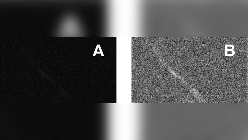
The results show that CARS readily visualizes cell membranes and lipid droplets, providing a morphological context for each cell. SHG images initially (day 0) display negligible signal for both cell types, confirming the low baseline collagen content. However, a faint SHG signal is already detectable in hCFs at day 0, indicating the system’s high sensitivity—collagen can be observed after only a few hours of culture. By day 7, both hCFs and hMSCs exhibit clear SHG contrast, with hCFs showing well‑defined collagen networks and hMSCs beginning to produce collagen, suggesting the onset of differentiation. Subsequent time points (days 14, 21, 28) reveal progressive collagen deposition throughout the cytoplasm of both cell types, while the nuclear region remains SHG‑dark, reinforcing that the signal originates from extracellular matrix protein rather than autofluorescence. The authors note that images were acquired from different regions and samples at each time point, so quantitative spatial comparisons are not presented; nevertheless, the consistent detection of collagen over four weeks demonstrates the method’s robustness.
Image processing of SHG data involves contrast enhancement, Gaussian blurring, and manual brightness/contrast adjustment in ImageJ, converting low‑contrast raw images into quasi‑binary maps that clearly delineate collagen fibers. The authors emphasize that the laser powers used did not affect cell viability or proliferation, as evidenced by normal growth patterns across the experimental period.
In the discussion, the authors compare their approach to conventional collagen assays (Western blot, immunofluorescence, gene expression analysis), highlighting that those techniques are destructive, require fixation or large sample amounts, and lose spatial information. In contrast, the CARS‑SHG platform provides simultaneous chemical specificity (lipids vs. collagen), three‑dimensional resolution, and the ability to monitor the same living construct over time without perturbation. The study also confirms that fibrin gel, even in the absence of exogenous biochemical cues, can promote MSC differentiation toward a collagen‑producing phenotype, underscoring its relevance as a biomimetic scaffold for tissue engineering.
The paper concludes that multimodal CARS‑SHG microscopy is a powerful, non‑invasive tool for longitudinal studies of extracellular matrix formation in 3‑D cultures. Its low detection limit, deep tissue penetration (up to ~4 mm), and label‑free operation make it attractive for regenerative medicine applications, scaffold optimization, and quality control of cell‑based therapies. Future work may extend this methodology to more complex organoid systems, in vivo imaging, or integration with additional nonlinear modalities (e.g., two‑photon fluorescence) to provide a comprehensive picture of cell behavior within engineered tissues.
Comments & Academic Discussion
Loading comments...
Leave a Comment