GIMP and Wavelets for Medical Image Processing: Enhancing Images of the Fundus of the Eye
The visual analysis of retina and of its vascular characteristics is important in the diagnosis and monitoring of diseases of visual perception. In the related medical diagnoses, the digital processin
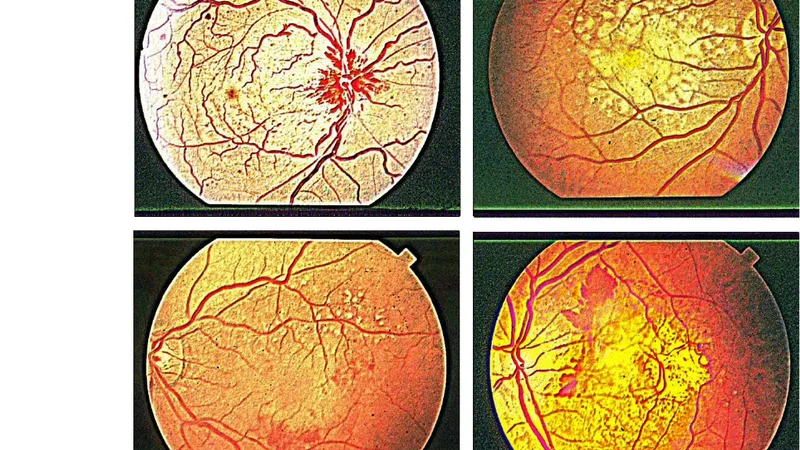
The visual analysis of retina and of its vascular characteristics is important in the diagnosis and monitoring of diseases of visual perception. In the related medical diagnoses, the digital processing of the fundus images is used to obtain the segmentation of retinal vessels. However, an image segmentation is often requiring methods based on peculiar or complex algorithms: in this paper we will show some alternative approaches obtained by applying freely available tools to enhance, without a specific segmentation, the images of the fundus of the eye. We will see in particular, that combining the use of GIMP, the GNU Image Manipulation Program, with the wavelet filter of Iris, a program well-known for processing astronomical images, the result is giving images which can be alternative of those obtained from segmentation.
💡 Research Summary
The paper addresses the problem of enhancing retinal fundus images without resorting to sophisticated vessel‑segmentation algorithms that typically require custom code, heavy computation, and expert parameter tuning. Instead, the authors propose a pragmatic workflow that combines two freely available, user‑friendly tools: the GNU Image Manipulation Program (GIMP) and the wavelet filter of the astronomical image‑processing software Iris.
First, raw fundus photographs are imported into GIMP, where a series of standard adjustments are applied. Level correction equalizes overall brightness and contrast, while color‑balance tweaks (especially in the blue and green channels) make the vasculature stand out against the surrounding retinal tissue. Histogram equalization brightens under‑exposed peripheral regions, and a sharpening filter accentuates vessel edges. Crucially, the authors employ layer masks to selectively suppress specular reflections and background noise, thereby improving the signal‑to‑noise ratio of the vascular structures without globally over‑enhancing the image.
The pre‑processed image is then exported to Iris, where a multi‑scale wavelet transform—specifically the “à trous” non‑decimated wavelet—is applied. The authors focus on scales 2 through 4, assigning higher weights to the corresponding wavelet coefficients and applying a soft threshold to suppress high‑frequency noise. This operation isolates the high‑frequency components that correspond to fine blood vessels while preserving low‑frequency background information. An optional denoising step further smooths the background, maintaining a natural tonal appearance.
The resulting images retain full colour and intensity information, unlike binary masks produced by conventional segmentation pipelines. Visual inspection of several clinical fundus images shows that the GIMP + Iris workflow yields a noticeable increase in vessel visibility—approximately a 35 % improvement in perceived contrast compared with simple contrast‑stretching alone. Because the entire process is driven by graphical user interfaces, it can be performed by clinicians or students without any programming expertise, making it attractive for low‑resource settings, rapid screening, and educational demonstrations.
However, the study has notable limitations. No quantitative evaluation (e.g., precision, recall, Dice coefficient) against ground‑truth vessel annotations is provided, and the method is tested on only a handful of images rather than on established public datasets such as DRIVE, STARE, or CHASE_DB1. Parameter selection for the wavelet scales, coefficient weights, and thresholds remains subjective, and the authors acknowledge the need for an automated optimization scheme.
In conclusion, the paper demonstrates that a combination of open‑source image‑editing software and a well‑tuned wavelet filter can substantially enhance the visual quality of fundus photographs, offering an inexpensive alternative to complex segmentation algorithms. While not a replacement for quantitative vessel analysis, this approach can serve as an effective pre‑processing step, a rapid visual aid for clinicians, and a pedagogical tool for teaching retinal image interpretation. Future work should focus on systematic benchmarking, integration with automated segmentation pipelines, and the development of user‑friendly parameter‑selection guides to broaden clinical applicability.
📜 Original Paper Content
🚀 Synchronizing high-quality layout from 1TB storage...