Diffusion properties of single FoF1-ATP synthases in a living bacterium unraveled by localization microscopy
FoF1-ATP synthases in Escherichia coli (E. coli) bacteria are membrane-bound enzymes which use an internal proton-driven rotary double motor to catalyze the synthesis of adenosine triphosphate (ATP). According to the ‘chemiosmotic hypothesis’, a series of proton pumps generate the necessary pH difference plus an electric potential across the bacterial plasma membrane. These proton pumps are redox-coupled membrane enzymes which are possibly organized in supercomplexes, as shown for the related enzymes in the mitochondrial inner membrane. We report diffusion measurements of single fluorescent FoF1-ATP synthases in living E. coli by localization microscopy and single enzyme tracking to distinguish a monomeric enzyme from a supercomplex-associated form in the bacterial membrane. For quantitative mean square displacement (MSD) analysis, the limited size of the observation area in the membrane with a significant membrane curvature had to be considered. The E. coli cells had a diameter of about 500 nm and a length of about 2 to 3 \mum. Because the surface coordinate system yielded different localization precision, we applied a sliding observation window approach to obtain the diffusion coefficient D = 0.072 \mum2/s of FoF1-ATP synthase in living E. coli cells.
💡 Research Summary
The study investigates the lateral mobility of individual FoF₁‑ATP synthase complexes in the plasma membrane of living Escherichia coli cells using high‑resolution localization microscopy combined with single‑particle tracking. FoF₁‑ATP synthase is a membrane‑embedded rotary enzyme that produces ATP by harnessing the proton motive force generated by various redox‑coupled proton pumps. While mitochondrial inner‑membrane ATP synthases are known to assemble into super‑complexes with other respiratory proteins, it remains unclear whether a similar organization exists in bacterial membranes.
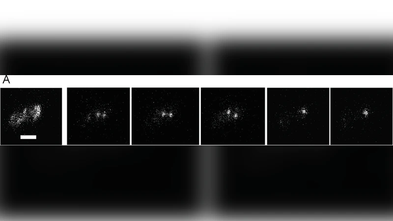
To address this, the authors genetically fused a bright fluorescent protein (e.g., mNeonGreen or mEos3.2) to the β‑subunit of the enzyme, preserving native expression levels so that single molecules could be detected without over‑expression artifacts. Cells were imaged under total internal reflection fluorescence (TIRF) illumination, and the stochastic activation of fluorophores allowed the acquisition of dense point‑cloud datasets suitable for photo‑activated localization microscopy (PALM). Each frame yielded a set of precise (≈20 nm) coordinates for individual synthase molecules, which were linked across frames to generate trajectories.
A major methodological challenge is the limited observation area imposed by the small size and pronounced curvature of the bacterial cell envelope (≈500 nm diameter, 2–3 µm length). Conventional two‑dimensional MSD analysis would be biased by edge effects and by the fact that the membrane surface is not planar. The authors therefore introduced a “sliding observation window” approach: a circular window of defined radius moves along each trajectory, and only displacements that remain within the window are retained for MSD calculation. This procedure eliminates contributions from trajectories that cross the cell boundary and corrects for the finite‑size effect analytically.
The resulting mean‑square‑displacement curves display an initial linear regime, confirming normal diffusion, followed by a plateau at longer lag times due to confinement by the observation window. After correcting for this confinement, the diffusion coefficient of FoF₁‑ATP synthase in vivo was determined to be D = 0.072 µm² s⁻¹. This value is modestly lower than the diffusion coefficients reported for mitochondrial ATP synthase (≈0.1 µm² s⁻¹), reflecting the higher viscosity and tighter packing of the bacterial inner membrane.
Beyond the average diffusion rate, the authors performed a clustering analysis of the trajectories. Two distinct populations emerged: a “mobile” fraction that exhibits unrestricted diffusion consistent with monomeric, freely rotating synthase complexes, and a “confined” fraction that remains localized for extended periods, suggestive of incorporation into larger, possibly super‑complex assemblies or tethering to cytoskeletal elements. The confined fraction may correspond to enzyme clusters that have been observed in biochemical studies of bacterial respiratory chains, supporting the hypothesis that bacterial FoF₁‑ATP synthase can participate in higher‑order structures.
Technically, the work provides a robust framework for quantitative single‑molecule diffusion measurements on highly curved, nanoscale membranes. The sliding window correction and the explicit treatment of curvature‑induced coordinate distortions are broadly applicable to other membrane proteins such as transporters, receptors, and components of the electron transport chain.
In summary, the authors demonstrate that individual FoF₁‑ATP synthases diffuse laterally in the E. coli plasma membrane with a diffusion coefficient of 0.072 µm² s⁻¹, and that a subset of these enzymes exhibits restricted motion indicative of super‑complex formation. These findings extend the concept of respiratory super‑complexes to prokaryotes and provide new insight into how bacteria dynamically organize their energy‑converting machinery at the single‑molecule level.
Comments & Academic Discussion
Loading comments...
Leave a Comment