What Brown saw and you can too
A discussion is given of Robert Brown’s original observations of particles ejected by pollen of the plant \textit{Clarkia pulchella} undergoing what is now called Brownian motion. We consider the nature of those particles, and how he misinterpreted the Airy disc of the smallest particles to be universal organic building blocks. Relevant qualitative and quantitative investigations with a modern microscope and with a “homemade” single lens microscope similar to Brown’s, are presented.
💡 Research Summary
The paper revisits Robert Brown’s 1827 observations of particles released from the pollen of Clarkia pulchella, which he famously described as “molecules” undergoing random motion—what we now call Brownian motion. By combining modern optical theory, electron microscopy, and a replica of Brown’s single‑lens “ball” microscope, the authors demonstrate that the smallest circular objects Brown reported were not actual organelles but diffraction artifacts (Airy discs) produced by his simple lens.
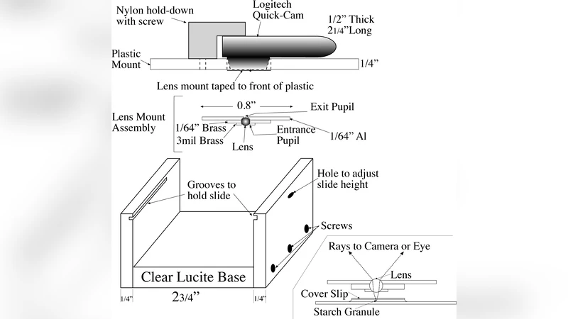
First, the authors reconstruct Brown’s microscope: a single spherical lens with a focal length of roughly 1/32 in (≈0.8 mm) giving a nominal magnification of about 320–370×. Using the same geometry, they show that the resolution limit is set by the Rayleigh criterion, and that objects smaller than ~1 µm appear as bright circular spots whose apparent diameter is dominated by the Airy pattern rather than the true particle size.
Second, the study employs a modern transmission electron microscope (TEM) and a high‑quality compound microscope to measure the true dimensions of the two main organelles found in Clarkia pollen: amyloplasts (starch‑storage bodies) and spherosomes (lipid‑storage bodies). The TEM data reveal diameters ranging from 0.5 µm to 1.5 µm for amyloplasts and 0.3 µm to 0.8 µm for spherosomes—significantly smaller than Brown’s estimate of 1.3–1.7 µm. The discrepancy is attributed to the diffraction‑limited image formed by his lens and to his method of comparing particle size against a micrometer slide with 5 µm divisions.
Third, the authors derive the expected mean‑square displacement of a particle undergoing Brownian motion in water at 20 °C using the Langevin‑Stokes framework. For a spherical particle of effective radius (R_{\rm eff}), the viscous drag coefficient is (\beta = 6\pi\eta R_{\rm eff}). The mean square displacement in one dimension is (\langle x^{2}\rangle = 2kTt/\beta). Table I in the paper translates this formula into concrete numbers: a 1 µm particle moves on average ~0.5 µm in 1 s, ~2.9 µm in 30 s, and ~4 µm in 60 s. By comparing these distances with the human eye’s angular resolution (≈2.9 × 10⁻⁴ rad) and the magnification of Brown’s microscope, the authors argue that only particles smaller than about 4 µm produce observable jitter within a one‑second interval. Larger pollen grains (~100 µm) would move far less than the eye can resolve, explaining why Brown never saw the pollen itself drift.
Fourth, the paper provides a step‑by‑step guide to building a low‑cost “ball‑lens” microscope that reproduces Brown’s optical configuration. A 2 mm glass sphere is mounted in a holder, spaced appropriately from the specimen plane to achieve the 1/32 in focal length. The resulting instrument reaches magnifications of 300–400×, sufficient to resolve the amyloplasts and spherosomes and to observe their Brownian jitter. The authors validate the homemade device by comparing images and motion statistics with those obtained from a commercial compound microscope.
Finally, the historical narrative is clarified. Brown’s claim that the pollen itself moved was a misinterpretation; the pollen grain (≈100 µm) is far too large for its Brownian motion to be visible. The “molecules” he described were actually the intracellular organelles that happen to be of a size that produces observable jitter under his modest magnification. Moreover, Brown’s early speculation that these particles represented universal organic building blocks was later abandoned after he observed similar jitter in crushed glass, rocks, and even a fragment of the Sphinx.
In summary, the paper re‑examines a classic scientific observation with modern tools, quantifies the optical and hydrodynamic limits that shaped Brown’s conclusions, and offers an inexpensive experimental platform for educators and hobbyists to replicate the original phenomenon. It bridges historical insight with contemporary physics, demonstrating how diffraction, viscous drag, and human visual thresholds together produced the iconic story of “Brownian motion.”
Comments & Academic Discussion
Loading comments...
Leave a Comment