Two-dimensional wave patterns of spreading depolarization: retracting, re-entrant, and stationary waves
We present spatio-temporal characteristics of spreading depolarizations (SD) in two experimental systems: retracting SD wave segments observed with intrinsic optical signals in chicken retina, and spontaneously occurring re-entrant SD waves that repeatedly spread across gyrencephalic feline cortex observed by laser speckle flowmetry. A mathematical framework of reaction-diffusion systems with augmented transmission capabilities is developed to explain the emergence and transitions between these patterns. Our prediction is that the observed patterns are reaction-diffusion patterns controlled and modulated by weak nonlocal coupling. The described spatio-temporal characteristics of SD are of important clinical relevance under conditions of migraine and stroke. In stroke, the emergence of re-entrant SD waves is believed to worsen outcome. In migraine, retracting SD wave segments cause neurological symptoms and transitions to stationary SD wave patterns may cause persistent symptoms without evidence from noninvasive imaging of infarction.
💡 Research Summary
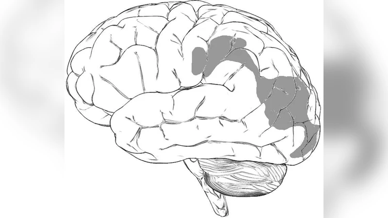
The paper investigates two‑dimensional spreading depolarization (SD) wave patterns in two experimental preparations: retracting SD wave segments recorded with intrinsic optical signals in the chicken retina, and spontaneously occurring re‑entrant SD waves that repeatedly circulate across the gyrencephalic feline cortex as measured by laser speckle flowmetry. In the retinal preparation, an initially circular depolarization front breaks up and the remaining fragment retreats, producing a “retracting” wave segment that terminates without leaving a permanent lesion. In the feline cortical preparation, the depolarization front does not extinguish; instead it becomes trapped by anatomical or functional heterogeneities (e.g., sulcal‑gyral geometry, vascular architecture) and forms a closed trajectory that repeatedly re‑excites the same cortical region, i.e., a “re‑entrant” wave.
To explain both phenomena within a unified theoretical framework, the authors extend the classic reaction‑diffusion (RD) description of SD by adding a weak non‑local coupling term. The standard RD equations capture local excitatory dynamics (ionic shifts, membrane depolarization) and diffusion of extracellular potassium and other ions. However, real brain tissue exhibits long‑range interactions mediated by electrical synapses, chemical diffusion through perivascular spaces, and neurovascular feedback that can either suppress or facilitate propagation over distances larger than the local diffusion length. The non‑local term is modeled as an integral kernel with a characteristic spatial scale and a coupling strength that can be either excitatory or inhibitory.
Numerical simulations of the augmented model reveal three distinct regimes depending on the strength of the non‑local inhibitory component: (1) weak inhibition allows the wave to close on itself, producing a stable re‑entrant orbit; (2) intermediate inhibition causes the wave front to lose coherence at its leading edge, generating retreating fragments that shrink and disappear; (3) strong inhibition arrests propagation altogether, yielding a stationary, non‑propagating depolarization plateau. The transitions among these regimes reproduce the experimentally observed shift from retracting to re‑entrant to stationary patterns.
Clinically, the authors argue that these pattern dynamics have direct relevance to migraine and stroke. In migraine aura, retracting wave segments correspond to transient neurological symptoms without detectable infarction; however, if a retracting wave transitions to a stationary pattern, prolonged depolarization may sustain metabolic stress and produce persistent aura‑like symptoms despite normal imaging. In acute ischemic stroke, re‑entrant SD waves are especially deleterious: each circuitous pass repeatedly imposes metabolic demand on already compromised tissue, exacerbates perfusion deficits, and expands the infarct core. Consequently, therapeutic strategies that modulate the weak non‑local coupling—such as pharmacological agents that enhance inhibitory neurovascular feedback, NMDA receptor antagonists, or low‑intensity direct current stimulation—could shift the system toward the retracting or fully arrested regime, thereby mitigating tissue damage.
In summary, the study combines high‑resolution optical imaging, laser speckle flowmetry, and a mathematically rigorous extension of reaction‑diffusion theory to demonstrate that SD waveforms are not monolithic but can adopt retracting, re‑entrant, or stationary configurations governed by weak non‑local interactions. This unified view provides a mechanistic basis for interpreting diverse SD‑related phenomena in neurological disease and suggests novel avenues for targeted intervention.
Comments & Academic Discussion
Loading comments...
Leave a Comment