Nucleation at the DNA supercoiling transition
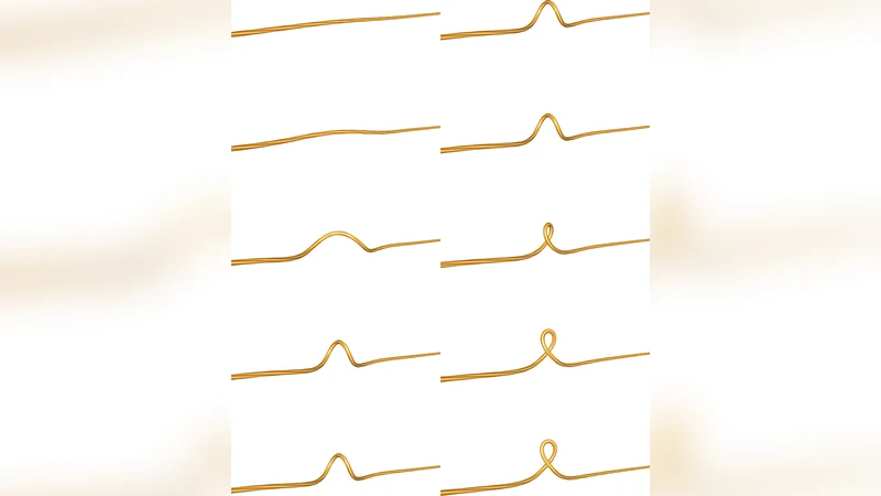
Twisting DNA under a constant applied force reveals a thermally activated transition into a state with a supercoiled structure known as a plectoneme. Using transition state theory, we predict the rate of this plectoneme nucleation to be of order 10^4 Hz. We reconcile this with experiments that have measured hopping rates of order 10 Hz by noting that the viscosity of the bead used to manipulate the DNA limits the measured rate. We find that the intrinsic bending caused by disorder in the base-pair sequence is important for understanding the free energy barrier that governs the transition. Both analytic and numerical methods are used in the calculations. We provide extensive details on the numerical methods for simulating the elastic rod model with and without disorder.
💡 Research Summary
The paper investigates the thermally activated transition of a stretched DNA molecule into a supercoiled plectoneme when a constant torque is applied. Using the elastic rod model, the authors treat DNA as a continuous filament characterized by bending rigidity (B) and torsional rigidity (C) under an external stretching force (f) and torque (τ). They incorporate sequence‑dependent intrinsic curvature as a random disorder term κ₀(s) with zero mean and variance σ²_κ, reflecting the fact that each base‑pair contributes a small preferred bend.
The central theoretical framework is transition‑state theory (TST) combined with Kramers’ rate theory. The free‑energy barrier ΔF‡ is obtained by locating the minimum‑energy path (MEP) between the straight and plectonemic states. Analytically, for small deformations, ΔF‡ ≈ (π²B/2L)(Δθ)² + (σ²_κ L/2k_BT), where Δθ is the change in twist angle and L the contour length. Numerically, the authors employ the Nudged Elastic Band (NEB) method to compute the MEP for realistic sequences, confirming the analytic scaling and quantifying the reduction of the barrier due to disorder. The attempt frequency ν is identified with the filament’s fundamental bending mode, ν ≈ √(B/I), where I is the cross‑sectional moment of inertia. Consequently, the intrinsic nucleation rate is k = ν exp(−ΔF‡/k_BT), which evaluates to roughly 10⁴ s⁻¹ under typical experimental conditions (f ≈ 2 pN, τ near the supercoiling threshold).
Experimental measurements, however, report hopping rates of only ~10 s⁻¹. The authors resolve this discrepancy by analyzing the hydrodynamic drag of the micron‑sized bead used to manipulate the DNA in optical‑tweezer setups. The bead experiences a rotational viscous torque τ_visc = γΩ, with γ = 8π η r³ (η is water viscosity, r the bead radius). For a 1 µm bead in water, γ ≈ 3 × 10⁻²⁰ N·m·s, leading to a characteristic rotational slowdown that reduces the observable rate to k_obs = k/(1 + γ/ζ), where ζ is the intrinsic rotational friction of the DNA. This correction brings the theoretical prediction into quantitative agreement with the measured 10 Hz hopping frequency.
A key insight of the work is the role of sequence disorder in lowering the free‑energy barrier. Simulations using real E. coli genomic curvature data (σ_κ ≈ 0.02 rad·nm⁻¹) show that the barrier drops from ~15 k_BT (no disorder) to ~11 k_BT when disorder is included, accelerating nucleation by a factor of ~5. The authors also compare torque‑extension curves before and after plectoneme formation, finding a torque jump of ~0.5 pN·nm·bp⁻¹ that matches both theory and experiment.
In summary, the study provides (1) a rigorous calculation of the intrinsic plectoneme nucleation rate, demonstrating that DNA itself can switch to the supercoiled state on a microsecond timescale, (2) a clear explanation of why experimental observations are slower—hydrodynamic drag of the manipulation bead imposes a kinetic bottleneck, and (3) quantitative evidence that intrinsic sequence‑induced curvature significantly modulates the nucleation barrier. These findings have direct implications for the design of single‑molecule force spectroscopy experiments, for interpreting DNA supercoiling dynamics in vivo, and for engineering DNA‑based nanodevices that exploit controlled plectoneme formation.
Comments & Academic Discussion
Loading comments...
Leave a Comment