Natural images from the birthplace of the human eye
Here we introduce a database of calibrated natural images publicly available through an easy-to-use web interface. Using a Nikon D70 digital SLR camera, we acquired about 5000 six-megapixel images of Okavango Delta of Botswana, a tropical savanna habitat similar to where the human eye is thought to have evolved. Some sequences of images were captured unsystematically while following a baboon troop, while others were designed to vary a single parameter such as aperture, object distance, time of day or position on the horizon. Images are available in the raw RGB format and in grayscale. Images are also available in units relevant to the physiology of human cone photoreceptors, where pixel values represent the expected number of photoisomerizations per second for cones sensitive to long (L), medium (M) and short (S) wavelengths. This database is distributed under a Creative Commons Attribution-Noncommercial Unported license to facilitate research in computer vision, psychophysics of perception, and visual neuroscience.
💡 Research Summary
The paper presents a rigorously calibrated natural‑image database collected in the Okavango Delta of Botswana, a tropical savanna environment thought to resemble the ecological niche in which the human visual system evolved. Using two Nikon D70 DSLR cameras equipped with an 18–70 mm f/3.5–4.5 lens and a 52 mm UV skylight filter, the authors acquired roughly 5,000 six‑megapixel photographs. The acquisition strategy combined opportunistic “baboon‑troop” sequences with controlled experiments that varied a single photographic parameter (aperture, object distance, time of day, horizon position).
A comprehensive camera characterization was performed. The angular resolution was measured to be 92 pixels per degree in both horizontal and vertical directions, slightly below the estimated 120 cones per degree of the human retina. Linearity of sensor response across a wide dynamic range was verified, and the consistency of shutter, aperture, and ISO settings was confirmed, enabling reliable conversion from camera settings to incident photon flux. Spectral sensitivities of the R, G, and B sensor channels were obtained by imaging a reflectance standard under 31 narrow‑band light sources; these measurements accurately predict sensor responses to broadband illumination. Dark current (sensor output with no light) and the spatial modulation transfer function (MTF) of each color plane were also quantified. Two separate D70 units yielded identical results apart from a single multiplicative scaling factor, which was corrected in the calibration pipeline.
With these measurements, raw camera RGB values were transformed into standardized R‑G‑B responses proportional to incident photon flux on each sensor type. The authors then converted these standardized values into physiologically relevant quantities for the three human cone classes (L, M, S). Using the Stockman‑Sharpe/CIE 2‑degree fundamentals and the method of Yin et al., they estimated cone isomerization rates (photoisomerizations · s⁻¹) for each pixel. In parallel, a grayscale luminance image was generated in units of cd · m⁻² based on the CIE 2007 photopic luminance function. Thus each photograph is available in four principal formats: (i) raw NEF (camera sensor output), (ii) demosaiced RGB MATLAB matrix, (iii) LMS MATLAB matrix (cone isomerization rates), and (iv) LUM MATLAB matrix (luminance). A fifth MATLAB structure supplies all metadata (exposure settings, calibration parameters, basic statistics).
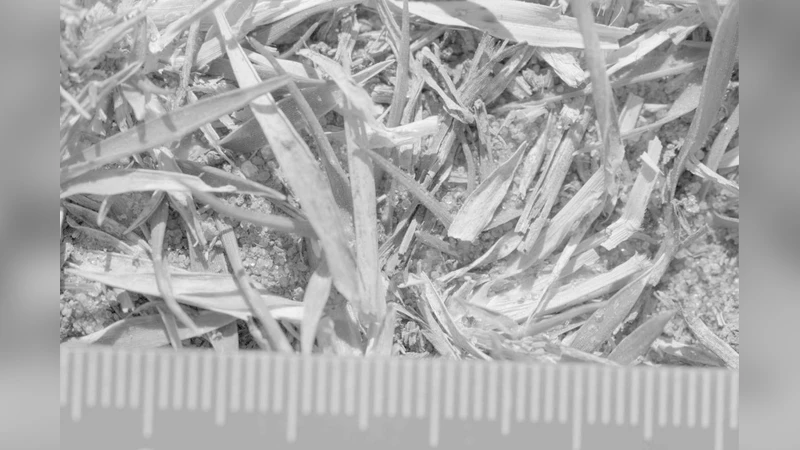
The dataset is organized into about 100 “albums,” each annotated with keywords and tags describing content (e.g., baboon habitat, sky‑horizon, ground close‑up) and methodological variables (e.g., fixed aperture, varying distance). Example analyses illustrate the database’s utility: (1) a time‑series of cloud‑free sky images taken every ten minutes from 06:30 to 18:30 shows diurnal changes in absolute luminance and relative L‑M‑S channel ratios, with the L channel becoming relatively stronger at sunrise and sunset; (2) a set of 23 close‑up grass images captured at different distances reveals that the pixel‑pixel luminance correlation function decays more rapidly for images taken from farther away, reflecting scale‑dependent texture statistics. These demonstrations underscore how the database can support investigations of spatial scaling, color statistics, and illumination dynamics in a biologically realistic setting.
Access to the full collection (≈300 GB across all formats) is provided via a web interface (http://tofu.physics.upenn.edu/~upennidb) and anonymous FTP (ftp://anonymous@tofu.physics.upenn.edu/fulldb). Users can select individual images or whole albums, choose desired output formats, and download recursively using tools such as wget. All content is released under a Creative Commons Attribution‑Noncommercial Unported license, permitting free non‑commercial use, redistribution, and remixing with appropriate attribution. Calibration software and detailed protocols are available on request.
In summary, this work delivers a uniquely valuable resource for vision science: a large‑scale, high‑resolution, physically calibrated natural‑image corpus expressed directly in the units of human photoreceptor activation. It bridges the gap between uncontrolled internet image collections and the stringent quantitative needs of computational neuroscience, psychophysics, and computer‑vision research, enabling rigorous testing of models of early visual processing, color perception, and image‑statistics‑based algorithms.
Comments & Academic Discussion
Loading comments...
Leave a Comment