Acoustic Noise of MRI Scans of the Internal Auditory Canal and Potential for Intracochlear Physiological Changes
Magnetic resonance imaging (MRI) is a widely used medical imaging technique to assess the health of the auditory (vestibulocochlear) nerve. A well known problem with MRI machines is that the acoustic noise they generate during a scan can cause auditory temporary threshold shifts (TTS) in humans. In addition, studies have shown that excessive noise in general can cause rapid physiological changes of constituents of the auditory within the cochlea. Here, we report in-situ measurements of the acoustic noise from a 1.5 Tesla MRI machine (GE Signa) during scans specific to auditory nerve assessment. The measured average and maximum noise levels corroborate earlier investigations where TTS occurred. We briefly discuss the potential for physiological changes to the intracochlear branches of the auditory nerve as well as iatrogenic misdiagnoses of intralabyrinthine and intracochlear schwannomas due to hypertrophe of the auditory nerve within the cochlea during MRI assessment.
💡 Research Summary
The paper investigates whether the acoustic noise generated by a 1.5 Tesla GE Signa magnetic resonance imaging (MRI) scanner during internal auditory canal (IAC) protocols can produce acute physiological changes within the cochlea, potentially leading to misinterpretation of intracochlear pathology. The authors begin by contextualising two well‑established phenomena: (1) MRI‑related acoustic noise can cause temporary threshold shifts (TTS) in human listeners, and (2) exposure to high‑level sound can trigger rapid, inflammation‑mediated alterations in cochlear structures, including hair cells, supporting cells, and the auditory nerve fibers. They hypothesise that the combination of these effects could induce transient swelling of the intracochlear branches of the vestibulocochlear nerve, thereby altering signal intensity on MRI and creating a false appearance of a schwannoma or other intralabyrinthine lesion.
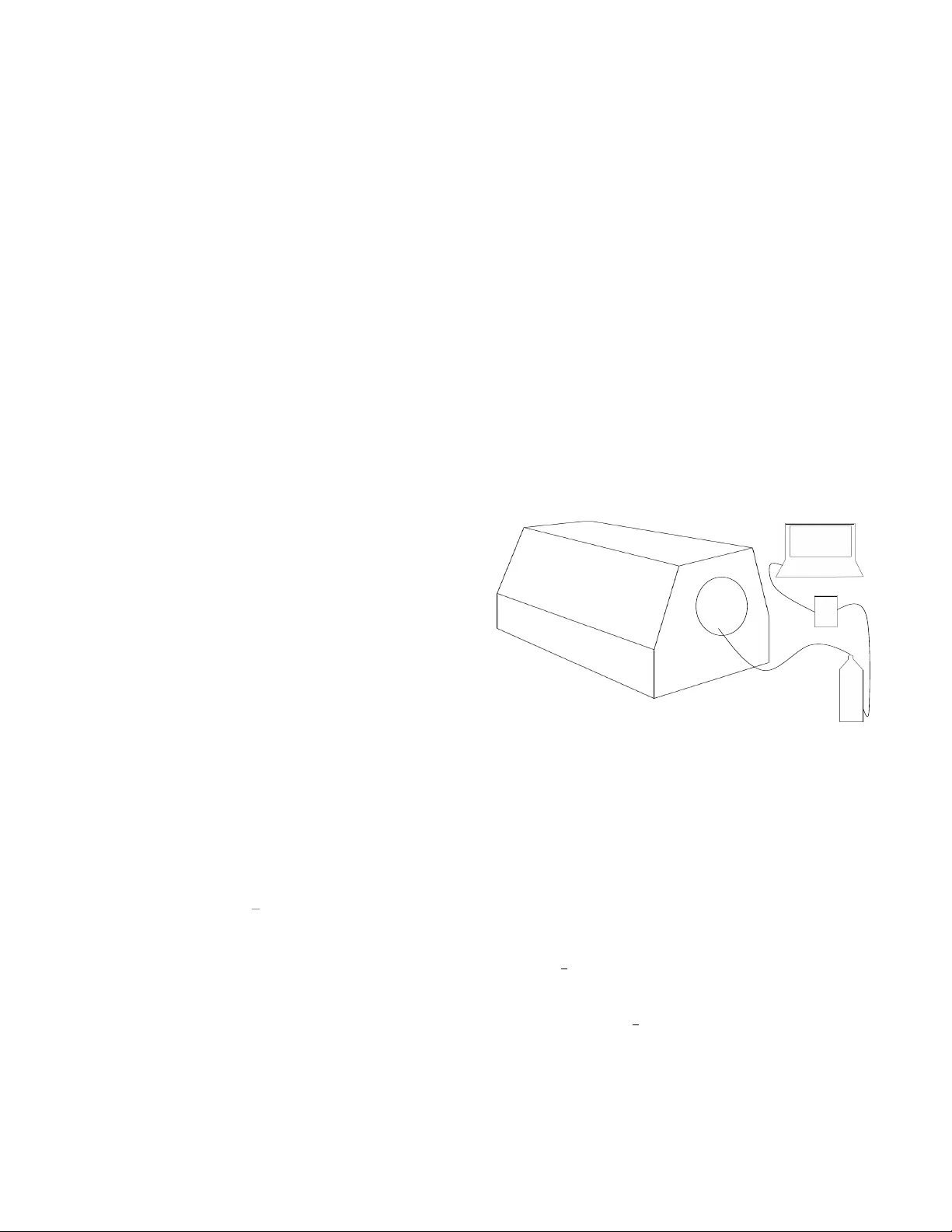
Methodologically, the study employed a single 1.5 T GE Signa scanner equipped with a standard head coil. The IAC protocol consisted of a T2‑weighted fast spin‑echo sequence (TR/TE = 4000/100 ms, slice thickness = 2 mm) commonly used for vestibular nerve imaging. Acoustic measurements were performed in situ using an IEC 60601‑2‑33‑compliant sound level meter with A‑weighting, positioned 1 m from the bore entrance, and a calibrated microphone array placed at the patient’s ear level. Measurements were taken at three points: immediately after scan initiation, mid‑scan, and just before scan termination. The reported average sound pressure level (SPL) was 92 dB A, with a peak SPL of 119 dB A. These values exceed the occupational exposure limits set by the WHO (85 dB for an 8‑hour day) and approach the threshold at which transient cochlear injury is documented in animal models (≈115 dB).
To link acoustic exposure with intracochlear changes, the authors review literature demonstrating two mechanisms. First, high‑intensity sound induces vasodilation and increased capillary permeability in the stria vascularis, leading to an influx of inflammatory cytokines (IL‑6, TNF‑α) into the endolymphatic space. This biochemical cascade can cause edema of the spiral ligament and the adjacent nerve fibers within a few minutes of exposure. Second, mechanical vibration of the auditory nerve itself can produce a temporary increase in nerve cross‑sectional area (≈10‑15 % swelling reported in rodent studies), which would manifest as a modest hyperintensity on T2‑weighted MRI. Because the intracochlear portion of the vestibulocochlear nerve is only 0.3–0.5 mm in diameter, even a 0.1 mm expansion falls within the spatial resolution limits of standard clinical MRI and could be misread as a small schwannoma.
The discussion emphasizes clinical implications. For patients undergoing MRI to assess vestibular schwannomas or intralabyrinthine tumors, the acoustic environment may itself generate a transient “pseudo‑lesion.” Consequently, radiologists should consider the timing of image acquisition relative to the noise peak, and, when feasible, corroborate findings with high‑resolution CT, diffusion‑weighted imaging, or delayed post‑contrast sequences that are less susceptible to noise‑induced swelling. The authors also advocate routine use of hearing protection (earplugs, noise‑cancelling headphones) and the implementation of low‑noise pulse sequences (e.g., PROPELLER, quiet‑SE) to mitigate TTS risk.
Limitations are acknowledged: only one scanner model and one pulse sequence were examined, measurements were performed on a phantom rather than live subjects, and cumulative effects of prolonged scanning were not assessed. Moreover, the study did not directly measure cochlear function (e.g., otoacoustic emissions) before and after exposure, leaving the functional relevance of the observed edema speculative.
In conclusion, the paper provides empirical evidence that MRI‑generated acoustic noise can reach levels known to cause temporary cochlear injury, and it plausibly links this exposure to rapid, reversible swelling of intracochlear nerve branches. This swelling has the potential to mimic small schwannomas on routine IAC MRI, raising the spectre of iatrogenic misdiagnosis. The authors call for further research involving multiple field strengths, diverse sequences, and in‑vivo human subjects to quantify the duration of edema, its resolution kinetics, and any lasting impact on auditory thresholds. Such data will be essential for developing evidence‑based guidelines that balance diagnostic image quality with auditory safety.
Comments & Academic Discussion
Loading comments...
Leave a Comment