A Quantum Mechanical Review of Magnetic Resonance Imaging
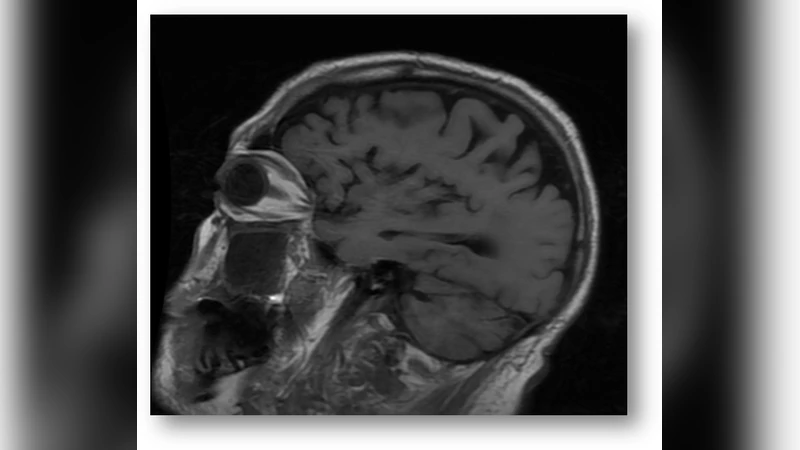
In this paper, we review the quantum mechanics of magnetic resonance imaging (MRI). We traverse its hierarchy of scales from the spin and orbital angular momentum of subatomic particles to the ensemble magnetization of tissue. And we review a number of modalities used in the assessment of acute ischemic stroke and traumatic brain injury.
💡 Research Summary
The paper provides a comprehensive quantum‑mechanical review of magnetic resonance imaging (MRI), tracing the physical principles from the subatomic level up to the macroscopic tissue response and then applying this framework to clinical modalities used for acute ischemic stroke and traumatic brain injury (TBI). It begins with a concise introduction that positions MRI as a cornerstone of modern diagnostic imaging while highlighting the inadequacy of purely classical descriptions for explaining many of its subtleties. The authors then lay out the quantum foundations: nuclear and electronic spin angular momentum, the associated magnetic moments, and the spin‑orbit coupling that distinguishes electron behavior from the proton (¹H) spins exploited in clinical MRI. The Larmor precession frequency ω₀ = γB₀ is derived from the Zeeman interaction, and the paper emphasizes that the gyromagnetic ratio γ varies among isotopes, thereby influencing resonance conditions.
Next, the manuscript bridges individual spin dynamics to ensemble magnetization. Using the density‑matrix formalism and the Markov master equation, the authors show how spin‑lattice (T₁) and spin‑spin (T₂) relaxation arise from spin‑phonon and spin‑spin interactions, respectively. They demonstrate the reduction of the many‑body quantum description to the phenomenological Bloch equations, explicitly linking the relaxation super‑operators to observable longitudinal and transverse magnetization components. The discussion includes temperature dependence, chemical shift, and J‑coupling effects, all of which are treated as perturbations to the Zeeman Hamiltonian.
The bulk of the paper is devoted to a systematic review of MRI pulse sequences and contrast mechanisms, each re‑interpreted through a quantum lens. Conventional T₁‑ and T₂‑weighted imaging is described as selective sampling of the longitudinal and transverse components of the ensemble density matrix after a radio‑frequency (RF) excitation. Diffusion‑weighted imaging (DWI) and diffusion‑tensor imaging (DTI) are presented as measurements of the phase dispersion caused by stochastic spin displacement, with the diffusion tensor emerging from an anisotropic relaxation operator that encodes tissue microstructure. Arterial spin labeling (ASL) is framed as a labeling of a sub‑ensemble of spins that travel with the blood flow, while blood‑oxygen‑level‑dependent (BOLD) contrast is modeled as a quantum exchange interaction between paramagnetic deoxy‑hemoglobin and surrounding water protons, leading to T₂* modulation.
Clinical applications are examined in depth. For acute ischemic stroke, the authors explain how abrupt reductions in cerebral blood flow cause a rapid rise in the apparent diffusion coefficient (ADC) and a concomitant increase in T₂* signal due to elevated deoxy‑hemoglobin. They quantify these changes using a spin‑diffusion model that incorporates blood‑brain barrier permeability and metabolic oxygen consumption. The paper also discusses reperfusion injury, illustrating how the restoration of oxygenated blood reverses the BOLD signal and can be captured with dynamic susceptibility contrast (DSC) techniques. In the context of TBI, the review covers micro‑hemorrhages, vasogenic edema, and neuronal membrane disruption. Each pathology produces distinct quantum signatures: micro‑bleeds alter the local magnetic susceptibility, leading to pronounced T₂* shortening; edema changes the water proton density and chemical shift distribution, affecting both T₁ and T₂; and membrane rupture modifies the exchange rates between intracellular and extracellular water, detectable with multi‑echo gradient‑echo sequences and quantitative susceptibility mapping.
The final section looks forward to emerging quantum technologies that could revolutionize MRI. The authors speculate on the integration of entangled photon sources for ultra‑low‑noise detection, superconducting quantum interference devices (SQUIDs) operating at higher fields, and quantum‑enhanced reconstruction algorithms that exploit quantum‑inspired priors. They argue that while current clinical scanners already embody quantum mechanics at their core, the next generation of “quantum‑MRI” systems may achieve sub‑millimeter resolution, real‑time functional imaging, and unprecedented sensitivity to metabolic changes.
In conclusion, the paper successfully unifies the microscopic quantum description of spin dynamics with the macroscopic imaging techniques used in modern neuro‑imaging. By doing so, it not only clarifies the physical origins of existing contrast mechanisms but also provides a solid theoretical platform for future innovations that could dramatically improve the diagnosis and monitoring of stroke, TBI, and other neurological disorders.