Probing biological light-harvesting phenomena by optical cavities
We propose a driven optical cavity quantum electrodynamics (QED) set up aimed at directly probing energy transport dynamics in photosynthetic biomolecules. We show that detailed information concerning energy transfer paths and delocalization of exciton states can be inferred (and exciton energies estimated) from the statistical properties of the emitted photons. This approach provides us with a novel spectroscopic tool for the interrogation of biological systems in terms of quantum optical phenomena which have been usually studied for atomic or solid-state systems, e.g. trapped atoms and semiconductor quantum dots.
💡 Research Summary
The authors present a novel spectroscopic scheme that merges cavity quantum electrodynamics (cavity QED) with photosynthetic light‑harvesting complexes, aiming to extract detailed information about excitonic energy transfer directly from the statistics of photons emitted by an optical cavity. The proposed setup consists of a sample of pigment‑protein complexes (specifically the Fenna‑Matthews‑Olson, or FMO, complex) placed inside a high‑Q optical cavity. The system is driven by an external laser (the pump) while the cavity mode itself acts as a continuous‑wave probe. By monitoring the mean photon number ⟨a†a⟩ and the second‑order correlation function g^(2)(0) of the light leaking from the cavity, the authors demonstrate that both static exciton energies and dynamic energy‑transfer pathways can be inferred without the need for conventional ultrafast multidimensional spectroscopy.
The theoretical model combines (i) the Frenkel‑exciton Hamiltonian for the seven bacteriochlorophyll sites of FMO, (ii) a single‑polarization cavity mode with resonance frequency ω_c and decay rate κ = ω_c/Q, (iii) a dipole‑type interaction H_c = ∑_i g μ_i(σ_i^+ a + σ_i^- a†) where g ∝ √(ω_c/2εV) and μ_i are the transition dipole projections, and (iv) a classical laser field coupling H_l(t) = −∑_i μ_i E(t) σ_i^+ + h.c. Environmental dephasing is introduced via a Lindblad Haken‑Strobl term with rate γ ranging from 1 to 100 cm⁻¹, covering realistic protein‑solvent fluctuations.
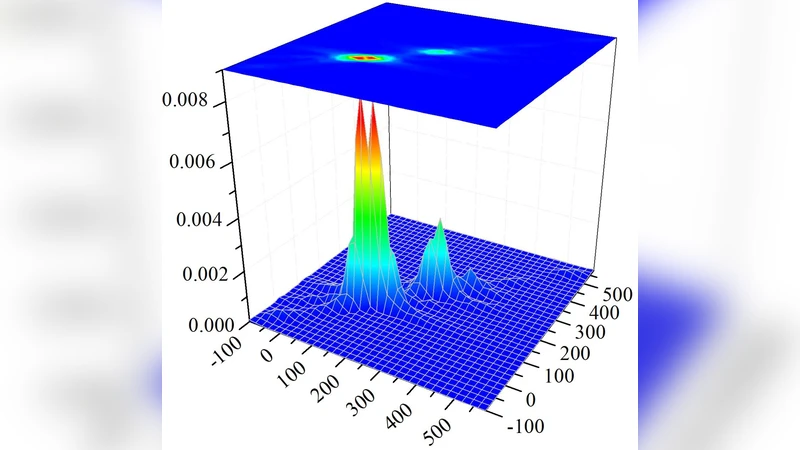
Numerical simulations of the full Liouville‑von Neumann master equation reveal a rich two‑dimensional landscape when ⟨a†a⟩ is plotted versus pump frequency ω_l and cavity frequency ω_c. Diagonal features (ω_l ≈ ω_c) align with the seven exciton energies of FMO, reproducing the familiar “peak‑on‑diagonal” pattern of 2D electronic spectroscopy. Off‑diagonal ridges appear where the cavity is resonant with one exciton while the pump addresses another, signalling coherent population transfer between exciton states. At higher pump intensities (≈ 10² kW cm⁻²) the diagonal peaks split, displaying an anti‑crossing reminiscent of dressed‑state (Rabi) splitting; this is interpreted as a laser‑induced hole‑burning effect that creates two new polaritonic branches.
Photon‑statistics analysis shows that, except on the exact diagonal (where g^(2)(0) ≈ 1, indicating a coherent state), the emitted light is sub‑Poissonian (g^(2)(0) < 1), i.e., anti‑bunched, revealing non‑classical photon generation driven by the coherent exciton dynamics. The degree of anti‑bunching is only weakly affected by dephasing, whereas the overall photon population drops sharply with increasing γ, suggesting that g^(2)(0) could serve as a robust probe of environmental noise strength.
To quantify quantum correlations between the cavity field and the biological sample, the authors compute the logarithmic negativity. Peaks in this entanglement measure appear around 800 fs after excitation and decay on a ~2 ps timescale, consistent with the fastest dephasing processes. This transient entanglement indicates the formation of polaritons—hybrid light‑matter quasiparticles—within a single FMO complex coupled to the cavity.
Experimental feasibility is addressed: state‑of‑the‑art photonic crystal or micro‑disk cavities can achieve Q ≈ 10⁴–10⁵ with mode volumes V ≈ 5 λ³, yielding coupling strengths g ≈ 0.015 cm⁻¹·D; optimistic designs approaching the diffraction limit (V ≈ (λ/2)³) could push g to ≈ 0.1 cm⁻¹·D, sufficient to observe the predicted effects. Random orientation averaging was included in the simulations, but the authors note that orientational ordering would enhance signal contrast.
In summary, the paper establishes that cavity‑QED spectroscopy can directly map excitonic energy landscapes and coherent transport pathways of photosynthetic complexes onto measurable photon observables. It simultaneously demonstrates the generation of non‑classical light, the emergence of polariton states, and the possibility of using photon‑statistics as a diagnostic of environmental decoherence. The approach offers a complementary, potentially less invasive alternative to ultrafast multidimensional spectroscopy and opens avenues for probing both natural and artificial light‑harvesting systems, as well as for engineering entangled polaritonic states across separate biological samples.
Comments & Academic Discussion
Loading comments...
Leave a Comment