Undulation Instability of Epithelial Tissues
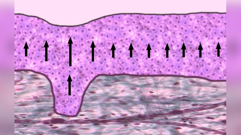
Treating the epithelium as an incompressible fluid adjacent to a viscoelastic stroma, we find a novel hydrodynamic instability that leads to the formation of protrusions of the epithelium into the stroma. This instability is a candidate for epithelial fingering observed in vivo. It occurs for sufficiently large viscosity, cell-division rate and thickness of the dividing region in the epithelium. Our work provides physical insight into a potential mechanism by which interfaces between epithelia and stromas undulate, and potentially by which tissue dysplasia leads to cancerous invasion.
💡 Research Summary
In this paper the authors investigate why the interface between an epithelial layer and its underlying stroma often displays undulating, finger‑like protrusions, especially in pre‑cancerous and cancerous tissues. Rather than invoking classical buckling or elastic growth models, they propose a new hydrodynamic instability that originates from the internal source of material—cell division—within the epithelium, which they treat as an incompressible viscous fluid. The stroma is modeled in two limiting cases: (i) a purely elastic solid characterized by a shear modulus µ, and (ii) a purely viscous fluid with viscosity ηₛ.
The governing equations for the epithelium consist of the incompressibility condition ∇·v = kₚ(z) and the Stokes‑type force balance η∇²v + η∇kₚ – ∇pₑ = 0, where the production rate kₚ(z)=k exp(‑z/l) − k₀ decays exponentially with distance from the interface, reflecting the reduced proliferative activity deeper in the tissue. The stroma obeys either µ∇²u − ∇pₛ = 0 (elastic) or ηₛ∇²vₛ − ∇pₛ = 0 (viscous). Boundary conditions enforce (a) Laplace pressure at the apical surface of the epithelium (surface tension γₐ), (b) zero displacement/velocity at the basal side of the stroma, (c) continuity of normal velocity and normal displacement at the epithelium‑stroma interface, (d) continuity of normal stress with an interfacial tension γᵢ, and (e) a tangential friction law ξ(Δvₓ)=0.
A flat base state is solved analytically, giving the vertical velocity profile v₀z(z) and pressure p₀e(z). The epithelium thickness H follows from the condition v₀z(H)=0. Small sinusoidal perturbations of the interface, δh(x,t)=δh₀ e^{ωt+iqx}, are introduced and the linearized equations are solved. The resulting eigenvalue problem yields three relaxation modes for an elastic stroma (ω_el₁, ω_el₂, ω_el₃) and two modes for a viscous stroma (ω_v₁, ω_v₂).
In the high‑wavenumber limit (q → ∞) the potentially unstable mode for the elastic case reads
ω_el₁ ≈ (η(k−k₀)/µ) − γᵢ q/(2η).
Similarly, for the viscous stroma the unstable mode is
ω_v₁ ≈ (η(k−k₀)/(η+ηₛ)) − γᵢ q/(2(η+ηₛ)).
Both expressions show that a sufficiently large product of viscosity η and net production (k−k₀) can overcome the stabilizing effects of interfacial tension γᵢ and the mechanical resistance of the stroma (µ or ηₛ). The other modes are always damped, being governed by surface tension at the apical surface or by tangential friction.
Parameter sweeps (Figures 2 and 3) demonstrate that increasing the epithelial viscosity η, the cell‑division rate k, the thickness of the proliferative zone l, or the stroma thickness L (elastic case) expands the region of positive ω, i.e., promotes instability. Conversely, larger shear modulus µ, larger stromal viscosity ηₛ, or higher interfacial tension γᵢ suppress the instability. The apical surface tension γₐ and the basal thickness L in the viscous case have only minor influence.
Biologically, the model captures several hallmarks of tumor progression: (1) malignant epithelia exhibit higher proliferation rates and a broader proliferative zone, both of which raise the term η(k−k₀) and thus the driving shear stress; (2) the surrounding stroma often becomes softer due to matrix remodeling and protease activity, effectively lowering µ or ηₛ and γᵢ, which further destabilizes the interface. The authors argue that protease‑mediated matrix degradation does not replace their mechanism but rather amplifies it by reducing the mechanical barriers. Consequently, the finger‑like invasion observed in dysplastic and cancerous tissues can be understood as a hydrodynamic amplification of a proliferative‑driven shear flow.
The paper also points out that the instability is generic to any viscous fluid with a volumetric source term; therefore, similar patterns could arise in engineered tissue constructs, bacterial colonies, or synthetic active fluids where material production creates internal flows. Experimental validation could be pursued in microfluidic systems where a controlled source of particles or cells is introduced into a viscous layer adjacent to a compliant substrate.
In summary, the authors identify a novel “production‑driven shear instability” at the epithelium‑stroma interface. The instability arises because cell division generates a depth‑dependent flow that creates shear stresses; when these stresses exceed the combined resistance of interfacial tension and stromal elasticity/viscosity, the flat interface becomes unstable and evolves into protruding fingers. This mechanism provides a physically grounded explanation for the enhanced undulations seen in early cancer invasion and suggests a broader class of active‑matter instabilities in biological and synthetic systems.
Comments & Academic Discussion
Loading comments...
Leave a Comment