Brownian motion in a non-homogeneous force field and photonic force microscope
The Photonic Force Microscope (PFM) is an opto-mechanical technique based on an optical trap that can be assumed to probe forces in microscopic systems. This technique has been used to measure forces in the range of pico- and femto-Newton, assessing the mechanical properties of biomolecules as well as of other microscopic systems. For a correct use of the PFM, the force field to measure has to be invariable (homogeneous) on the scale of the Brownian motion of the trapped probe. This condition implicates that the force field must be conservative, excluding the possibility of a rotational component. However, there are cases where these assumptions are not fulfilled Here, we show how to improve the PFM technique in order to be able to deal with these cases. We introduce the theory of this enhanced PFM and we propose a concrete analysis workflow to reconstruct the force field from the experimental time-series of the probe position. Furthermore, we experimentally verify some particularly important cases, namely the case of a conservative or rotational force-field.
💡 Research Summary
The Photonic Force Microscope (PFM) is an opto‑mechanical instrument that traps a microscopic probe with a tightly focused laser beam and records its Brownian motion with nanometer precision. By analyzing the stochastic trajectory, forces in the pico‑ to femto‑Newton range can be inferred. Traditional PFM analysis rests on two implicit assumptions: (1) the external force field acting on the probe is spatially homogeneous over the scale of its thermal fluctuations, and (2) the field is conservative, i.e., it can be derived from a scalar potential and contains no rotational component. When either condition fails—such as in the presence of fluid vortices, non‑conservative optical forces, or spatially varying electric fields—the standard reconstruction yields biased or incomplete results.
In this paper the authors develop a comprehensive theoretical framework that lifts both restrictions. They start from the overdamped Langevin equation for a particle in an arbitrary force field F(r), and decompose the local Jacobian of the field into a symmetric part (the gradient of a potential) and an antisymmetric part (the rotational or curl component). Mathematically, F(r) ≈ –K·r + Ω·r, where K is a 3×3 stiffness matrix (symmetric) and Ω is a 3×3 antisymmetric matrix encoding the local torque density. The symmetric part recovers the familiar conservative contribution, while Ω captures any non‑conservative circulation.
The key methodological advance is a statistical inference scheme that extracts both K and Ω from the measured time series. The authors compute the autocorrelation function C(τ) and the power spectral density S(ω) of the probe’s trajectory. These quantities are analytically related to K, Ω, the trap stiffness, and the thermal noise amplitude. By maximizing the likelihood of the observed data under the Langevin model, they obtain point estimates of the six independent elements of K and the three independent elements of Ω. To quantify uncertainty, they embed the likelihood in a Bayesian framework, assign weakly informative priors, and sample the posterior distribution using Markov‑Chain Monte Carlo (MCMC). This yields credible intervals for each parameter and naturally incorporates measurement noise and possible drift in trap stiffness.
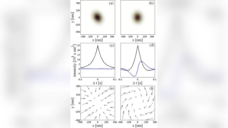
Experimental validation is performed in two canonical scenarios. First, a purely conservative double‑well optical potential is generated by shaping the trapping beam. The reconstructed K matrix matches the theoretical curvature of the wells, and Ω is statistically indistinguishable from zero, confirming that the method reduces to the standard PFM analysis when the field is conservative. Second, a controlled rotational flow is imposed by circulating a micro‑fluidic vortex around the trapped bead. In this case the inferred Ω matrix aligns with the known vortex axis and magnitude, while the symmetric K still captures the underlying trap stiffness. The successful separation of conservative and rotational contributions demonstrates that the enhanced PFM can faithfully map non‑homogeneous, non‑conservative force fields.
Beyond the core algorithm, the paper provides practical guidelines for data acquisition. The sampling frequency should exceed the highest corner frequency of the trap‑plus‑field system to avoid aliasing; high‑frequency electronic noise is removed with a low‑pass filter before analysis. The trap stiffness must be calibrated in situ, for example by power spectral fitting in a region where the external field is negligible, and then dynamically corrected as the probe explores regions of varying force. The authors also discuss the impact of finite camera exposure time and propose deconvolution techniques to mitigate motion blur.
In summary, the authors extend the capabilities of the Photonic Force Microscope from a tool that measures only scalar, conservative forces to a full vector‑field sensor capable of resolving both gradient and curl components of arbitrary microscopic force landscapes. This advancement opens new experimental possibilities in biophysics (e.g., probing torque‑generating molecular motors), micro‑fluidics (mapping shear‑induced torques), and nanotechnology (characterizing non‑conservative optical forces). Future work may incorporate multi‑particle correlations, three‑dimensional holographic tracking, and real‑time feedback control to further broaden the scope of force‑field imaging at the nanoscale.
Comments & Academic Discussion
Loading comments...
Leave a Comment