A preliminary study of acoustic propagation in thick foam tissue scaffolds composed of poly(lactic-co-glycolic acid)
The exclusive ability of acoustic waves to probe the structural, mechanical and fluidic properties of foams may offer novel approaches to characterise the porous scaffolds employed in tissue engineering. Motivated by this we conduct a preliminary investigation into the acoustic properties of a typical biopolymer and the feasibility of acoustic propagation within a foam scaffold thereof. Focussing on poly(lactic-co-glycolic acid), we use a pulse-echo method to determine the longitudinal speed of sound, whose temperature-dependence reveals the glass transition of the polymer. Finally, we demonstrate the first topographic and tomographic acoustic images of polymer foam tissue scaffolds.
💡 Research Summary
This paper presents a pioneering investigation into the acoustic properties of thick poly(lactic‑co‑glycolic acid) (PLGA) foam scaffolds, which are widely employed as porous matrices in tissue engineering. The authors argue that acoustic waves, unlike conventional imaging or mechanical testing techniques, can simultaneously probe structural, mechanical, and fluidic characteristics of foams in a non‑destructive manner, offering a potentially transformative tool for scaffold characterization.
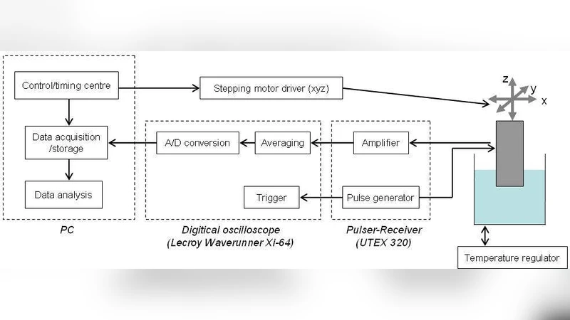
The experimental work began with the fabrication of PLGA 85:15 foams using a freeze‑drying method that produced scaffolds 2–3 mm thick, with an average pore diameter of ~150 µm and a porosity of ~85 %. The scaffolds were immersed in a temperature‑controlled water bath, and a single‑pulse, pulse‑echo ultrasound system (5 MHz center frequency) was used to acquire reflected signals from the scaffold front and back surfaces. By measuring the round‑trip travel time (Δt) and knowing the physical thickness (d) measured with a digital caliper, the longitudinal speed of sound (c = 2d/Δt) was calculated for each temperature.
Temperature‑dependent measurements were performed from 20 °C to 80 °C in 5 °C increments, with three repetitions per point. The data revealed a modest decrease in sound speed (≈2 %) between 20 °C and 40 °C, followed by a pronounced drop (≈6 %) between 45 °C and 55 °C. This abrupt change coincides with the glass‑transition temperature (Tg) of PLGA as determined by differential scanning calorimetry, confirming that ultrasonic velocity is highly sensitive to the polymer’s segmental mobility and can serve as a non‑invasive indicator of Tg.
Beyond bulk velocity, the authors demonstrated two acoustic imaging modalities. First, a surface topography map was generated from the latency of the first echo, achieving sub‑30 µm resolution of surface pore depth and morphology. Second, a tomographic reconstruction was obtained by laterally scanning the transducer while recording successive echoes, producing cross‑sectional images that reveal internal pore continuity, the ratio of closed‑to‑open pores, and the distribution of water within the scaffold. Notably, water‑filled pores caused a ~5 % reduction in local sound speed and an increase in attenuation, indicating that acoustic parameters can be used to quantify fluid content in situ.
The discussion emphasizes several strengths of the approach: (1) the ability to detect Tg without destroying the scaffold, enabling real‑time monitoring of thermal processing or storage conditions; (2) the low cost, rapid acquisition, and absence of ionizing radiation compared with X‑ray computed tomography; and (3) the feasibility of probing relatively thick scaffolds (several millimeters) with sufficient signal‑to‑noise ratio. Limitations are also acknowledged. The 5 MHz frequency is comparable to the characteristic pore size, leading to scattering‑induced resolution loss, and higher frequencies suffer greater intrinsic polymer attenuation, reducing penetration depth. Temperature control precision and potential thermal gradients in the water bath could introduce uncertainties in Tg estimation.
Future directions proposed include employing multi‑frequency ultrasound (1–10 MHz) to balance resolution and penetration, integrating ultrasonic photon‑correlation techniques for enhanced contrast, and expanding the methodology to other biodegradable polymers, composite scaffolds, and cell‑seeded constructs. The ultimate goal is to build a comprehensive acoustic property database that can be leveraged for quality control, design optimization, and real‑time, bedside monitoring of scaffold performance in regenerative medicine applications.
In conclusion, the study validates pulse‑echo ultrasound as a versatile, non‑destructive platform for measuring longitudinal sound speed, detecting the glass transition, and generating both surface and volumetric acoustic images of thick PLGA foam scaffolds. These capabilities open new avenues for scaffold assessment, offering a rapid, inexpensive, and clinically translatable alternative to existing characterization techniques.
Comments & Academic Discussion
Loading comments...
Leave a Comment