Nonparametric tests of structure for high angular resolution diffusion imaging in Q-space
High angular resolution diffusion imaging data is the observed characteristic function for the local diffusion of water molecules in tissue. This data is used to infer structural information in brain imaging. Nonparametric scalar measures are proposed to summarize such data, and to locally characterize spatial features of the diffusion probability density function (PDF), relying on the geometry of the characteristic function. Summary statistics are defined so that their distributions are, to first-order, both independent of nuisance parameters and also analytically tractable. The dominant direction of the diffusion at a spatial location (voxel) is determined, and a new set of axes are introduced in Fourier space. Variation quantified in these axes determines the local spatial properties of the diffusion density. Nonparametric hypothesis tests for determining whether the diffusion is unimodal, isotropic or multi-modal are proposed. More subtle characteristics of white-matter microstructure, such as the degree of anisotropy of the PDF and symmetry compared with a variety of asymmetric PDF alternatives, may be ascertained directly in the Fourier domain without parametric assumptions on the form of the diffusion PDF. We simulate a set of diffusion processes and characterize their local properties using the newly introduced summaries. We show how complex white-matter structures across multiple voxels exhibit clear ellipsoidal and asymmetric structure in simulation, and assess the performance of the statistics in clinically-acquired magnetic resonance imaging data.
💡 Research Summary
This paper introduces a fully non‑parametric framework for characterizing the micro‑structural geometry of brain white matter directly from high‑angular‑resolution diffusion imaging (HARDI) data. The authors observe that the measured HARDI signal at each voxel is mathematically the characteristic function (CF) of the underlying diffusion probability density function (PDF). Rather than inverting the CF or fitting parametric models such as multi‑tensor, Q‑ball, or spherical deconvolution, they exploit the geometry of the CF itself to construct scalar summary statistics that are analytically tractable and, to first order, independent of nuisance parameters such as signal‑to‑noise ratio (SNR).
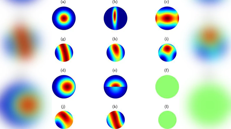
The methodology proceeds in three stages. First, the direction in which the CF attains its maximum is identified and taken as the dominant diffusion direction. Using this direction as a reference, a new orthogonal coordinate system (u₁, u₂, u₃) is defined in Fourier space: u₁ aligns with the dominant direction, while u₂ and u₃ span the orthogonal plane. Second, moments of the CF are computed in this basis. The second‑order moments (variances) quantify anisotropy, whereas higher‑order moments (skewness, kurtosis) capture asymmetry and multimodality. Crucially, each moment is normalized so that its sampling distribution under the null hypothesis follows a known parametric form (χ² or F), rendering the test statistics analytically derivable without resampling. Third, a hierarchy of hypothesis tests is applied: (i) isotropy versus anisotropy, (ii) unimodality versus multimodality, and (iii) symmetry versus asymmetry. Each test yields a p‑value, allowing the researcher to decide, at a pre‑specified significance level, whether the diffusion at a voxel exhibits a single dominant fiber, crossing fibers, or more exotic non‑Gaussian features.
To validate the approach, the authors conduct two complementary experiments. In simulation, they generate synthetic diffusion PDFs ranging from a single Gaussian (single fiber) to a mixture of two Gaussians (crossing fibers) and to asymmetric Lévy‑stable distributions. For each synthetic case, the proposed statistics correctly identify the underlying structure: the anisotropy measure distinguishes isotropic from anisotropic diffusion; the multimodality test reliably detects two peaks when the angular separation exceeds ~30°, and the asymmetry test flags Lévy‑stable PDFs with non‑zero skewness. In a clinical dataset acquired on a 3 T scanner, the method is applied voxel‑wise across the whole brain. Compared with conventional diffusion tensor imaging (DTI) metrics such as fractional anisotropy (FA) and mean diffusivity (MD), the non‑parametric tests reveal finer distinctions. In regions of known fiber crossing (e.g., the centrum semiovale), the multimodality test is strongly positive even when FA is low, indicating that the loss of anisotropy in DTI does not imply a lack of structural information. Moreover, in curved tracts and regions adjacent to ventricles, the asymmetry test identifies deviations from Gaussian symmetry that are invisible to DTI.
The authors discuss limitations and future extensions. Because the current implementation treats each voxel independently, spatial regularization or multivariate extensions could improve robustness, especially in low‑SNR regimes where the first‑order independence assumption weakens. They also note that higher‑order corrections or bootstrap refinements may be required for very noisy data. Nonetheless, the work demonstrates that operating directly in the Fourier domain provides a powerful, model‑free avenue for probing white‑matter architecture. By delivering analytically tractable, hypothesis‑driven statistics, the framework promises to complement existing diffusion analyses and to become a new standard for assessing complex microstructural features such as fiber crossings, dispersion, and non‑Gaussian asymmetry without imposing restrictive parametric forms.
Comments & Academic Discussion
Loading comments...
Leave a Comment