How can we kill cancer cells: insights from the computational models of apoptosis
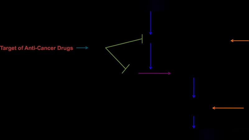
Cancer cells are widely known to be protected from apoptosis, which is a major hurdle to successful anti-cancer therapy. Over-expression of several anti-apoptotic proteins, or mutations in pro-apoptotic factors, has been recognized to confer such resistance. Development of new experimental strategies, such as in silico modeling of biological pathways, can increase our understanding of how abnormal regulation of apoptotic pathway in cancer cells can lead to tumour chemoresistance. Monte Carlo simulations are in particular well suited to study inherent variability, such as spatial heterogeneity and cell-to-cell variations in signaling reactions. Using this approach, often in combination with experimental validation of the computational model, we observed that large cell-to-cell variability could explain the kinetics of apoptosis, which depends on the type of pathway and the strength of stress stimuli. Most importantly, Monte Carlo simulations of apoptotic signaling provides unexpected insights into the mechanisms of fractional cell killing induced by apoptosis-inducing agents, showing that not only variation in protein levels, but also inherent stochastic variability in signaling reactions, can lead to survival of a fraction of treated cancer cells.
💡 Research Summary
The manuscript tackles a central problem in oncology: why many anti‑cancer agents fail to eradicate an entire tumor mass, leaving a resistant sub‑population behind. The authors argue that the conventional view—focused mainly on average expression levels of anti‑apoptotic proteins such as Bcl‑2, Bcl‑XL, or Mcl‑1—does not fully explain the phenomenon of “fractional killing.” To address this gap, they develop a stochastic, agent‑based Monte Carlo model of the two canonical apoptosis pathways (extrinsic/Fas‑TRAIL and intrinsic/mitochondrial) and couple it with experimental validation in human cancer cell lines.
Key methodological steps include: (1) defining a reaction network that captures ligand‑receptor binding, DISC formation, caspase activation, mitochondrial outer‑membrane permeabilization (MOMP), and downstream execution; (2) assigning probabilistic transition rates to each elementary step based on kinetic data from the literature and calibrated against dose‑response curves; (3) initializing each simulated cell with protein concentrations drawn from experimentally measured distributions, thereby embedding cell‑to‑cell heterogeneity; and (4) incorporating spatial constraints by modeling diffusion on a lattice, which allows the model to capture subcellular compartmentalization and local concentration fluctuations.
The simulations reveal two complementary sources of variability that shape the overall killing efficiency. First, the strength of the apoptotic stimulus determines the width of the death‑time distribution. Weak stimuli produce a broad spread, generating a pronounced “survival tail” where a fraction of cells never reaches the apoptotic threshold within the observation window. Second, even under strong stimuli, the intrinsic stochasticity of molecular interactions—particularly the formation of the apoptosome and the activation of executioner caspases—creates a non‑zero probability that a cell will escape death despite having a pro‑apoptotic protein profile that would be considered sufficient in deterministic models. This stochastic escape is amplified when anti‑apoptotic proteins are over‑expressed, but it is not eliminated; the model predicts survival rates of 15–30 % even in highly sensitized scenarios.
Experimental validation was performed using MCF‑7 (breast) and A549 (lung) cancer cells treated with varying concentrations of all‑trans retinoic acid, BH3‑mimetic peptides, and Fas ligand. Single‑cell death kinetics were recorded by time‑lapse fluorescence microscopy and flow cytometry. The empirical death‑time histograms matched the Monte Carlo predictions closely, confirming that the model captures both the mean response and the variance observed in real cell populations. Notably, the “survival tail” seen at low drug doses was reproduced without any ad‑hoc parameter tuning, supporting the authors’ claim that stochastic reaction dynamics are sufficient to generate fractional killing.
In the discussion, the authors translate these mechanistic insights into therapeutic implications. Traditional drug development focuses on achieving a target EC50 or maximizing overall tumor shrinkage, but the stochastic model suggests that eliminating the survival tail requires either (a) increasing the stimulus strength beyond a stochastic threshold (which may be toxic to normal tissue) or (b) simultaneously targeting multiple nodes of the apoptotic network to reduce the number of independent stochastic events that can rescue a cell. They illustrate this with a simulated combination of a Bcl‑2 inhibitor (e.g., venetoclax) and an Mcl‑1 inhibitor (e.g., AMG‑176), which synergistically compresses the death‑time distribution and drives predicted killing rates above 95 %. The authors propose that patient‑specific proteomic profiling could feed into the Monte Carlo framework to generate personalized drug‑combination recommendations—a true “in silico” precision‑medicine platform.
The conclusion emphasizes that Monte Carlo simulations of apoptosis provide a quantitative bridge between molecular heterogeneity and population‑level treatment outcomes. By accounting for both deterministic differences in protein abundance and the inherent randomness of biochemical reactions, the model explains why a fraction of cancer cells survive even the most potent pro‑apoptotic agents. Future work is outlined to extend the model to include immune‑cell interactions, tumor microenvironmental factors (hypoxia, nutrient gradients), and genomic mutations that alter reaction rates. Such extensions would further enhance the model’s realism and its utility in designing combination therapies that minimize fractional killing, ultimately improving clinical response durability.
Comments & Academic Discussion
Loading comments...
Leave a Comment