Classification of interstitial lung disease patterns with topological texture features
Topological texture features were compared in their ability to classify morphological patterns known as ‘honeycombing’ that are considered indicative for the presence of fibrotic interstitial lung diseases in high-resolution computed tomography (HRCT) images. For 14 patients with known occurrence of honey-combing, a stack of 70 axial, lung kernel reconstructed images were acquired from HRCT chest exams. A set of 241 regions of interest of both healthy and pathological (89) lung tissue were identified by an experienced radiologist. Texture features were extracted using six properties calculated from gray-level co-occurrence matrices (GLCM), Minkowski Dimensions (MDs), and three Minkowski Functionals (MFs, e.g. MF.euler). A k-nearest-neighbor (k-NN) classifier and a Multilayer Radial Basis Functions Network (RBFN) were optimized in a 10-fold cross-validation for each texture vector, and the classification accuracy was calculated on independent test sets as a quantitative measure of automated tissue characterization. A Wilcoxon signed-rank test was used to compare two accuracy distributions and the significance thresholds were adjusted for multiple comparisons by the Bonferroni correction. The best classification results were obtained by the MF features, which performed significantly better than all the standard GLCM and MD features (p < 0.005) for both classifiers. The highest accuracy was found for MF.euler (97.5%, 96.6%; for the k-NN and RBFN classifier, respectively). The best standard texture features were the GLCM features ‘homogeneity’ (91.8%, 87.2%) and ‘absolute value’ (90.2%, 88.5%). The results indicate that advanced topological texture features can provide superior classification performance in computer-assisted diagnosis of interstitial lung diseases when compared to standard texture analysis methods.
💡 Research Summary
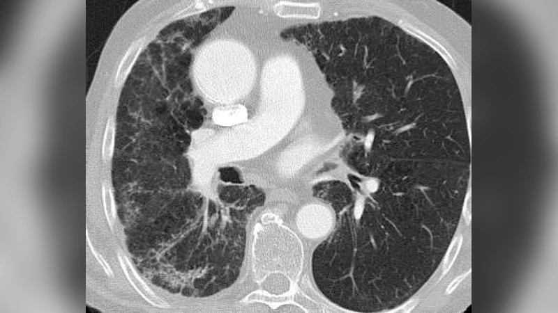
Interstitial lung diseases (ILD) are a heterogeneous group of disorders characterized by progressive fibrosis and loss of lung function. Early and accurate detection of fibrotic patterns on high‑resolution computed tomography (HRCT) is essential for patient management, yet conventional visual assessment remains subjective. This study investigates whether advanced topological texture descriptors—specifically Minkowski Functionals (MF)—can outperform traditional gray‑level co‑occurrence matrix (GLCM) features and Minkowski Dimension (MD) in automatically classifying the “honeycombing” pattern, a hallmark of advanced fibrosis.
Materials and Methods
Fourteen patients with confirmed honeycombing on HRCT were retrospectively selected. From each examination, a stack of 70 axial, lung‑kernel reconstructed slices was obtained. An experienced thoracic radiologist delineated 241 regions of interest (ROIs): 152 from apparently healthy lung parenchyma and 89 from pathological honeycombing zones. Each ROI measured 32 × 32 pixels and was intensity‑normalized prior to feature extraction.
Three families of texture features were computed:
- GLCM Features – Six conventional statistics (homogeneity, absolute value, contrast, energy, correlation, entropy) derived from 256 gray‑level co‑occurrence matrices.
- Minkowski Dimension (MD) – A fractal‑based estimate of image complexity obtained via box‑counting across multiple scales.
- Minkowski Functionals (MF) – Three topological measures (area, perimeter, Euler characteristic χ) calculated after binarizing each ROI at eight equally spaced thresholds. The Euler characteristic, χ, captures the number of connected components minus the number of holes, thereby encoding the intricate network‑like architecture of honeycombing.
For each ROI, the corresponding feature vector (GLCM, MD, or MF) was assembled. Two classifiers were trained and evaluated:
- k‑Nearest Neighbor (k‑NN) – Euclidean distance, with the optimal k (1–15) selected via 10‑fold cross‑validation.
- Multilayer Radial Basis Function Network (RBFN) – A feed‑forward neural network with a single hidden layer of radial basis units; the number of hidden nodes and Gaussian width were tuned through grid search within the same cross‑validation framework.
Performance was measured as classification accuracy on the held‑out test folds. To compare accuracy distributions, the Wilcoxon signed‑rank test was applied, and p‑values were Bonferroni‑corrected for multiple pairwise comparisons.
Results
The MF‑Euler characteristic (MF.euler) achieved the highest accuracies: 97.5 % with k‑NN and 96.6 % with RBFN. In contrast, the best conventional GLCM features were homogeneity (91.8 %/87.2 % for k‑NN/RBFN) and absolute value (90.2 %/88.5 %). MD alone yielded accuracies below 80 %. Statistical testing confirmed that MF.euler outperformed every GLCM and MD feature (p < 0.005 after Bonferroni correction). Both classifiers exhibited consistent superiority when using MF descriptors, indicating that the advantage is not classifier‑specific.
Discussion
The Euler characteristic directly quantifies the topological complexity of the honeycomb architecture—namely, the presence of multiple cystic air‑spaces surrounded by fibrotic walls. This property appears to be more discriminative than second‑order gray‑level statistics, which primarily capture local intensity relationships but ignore global connectivity. The findings suggest that incorporating topological information can substantially boost computer‑assisted diagnosis (CAD) of ILD. However, the study has notable limitations: ROIs were manually placed by a single radiologist, the sample size (14 patients) is modest, and all data originated from a single imaging protocol. Consequently, external validation on larger, multi‑center cohorts and automated ROI segmentation are required before clinical deployment.
Conclusion
Advanced topological texture analysis, particularly the Euler characteristic derived from Minkowski Functionals, provides a markedly superior means of distinguishing honeycombing from normal lung tissue on HRCT compared with standard GLCM or fractal‑dimension features. This work underscores the potential of topological descriptors to enhance CAD systems for interstitial lung disease, paving the way for more objective, reproducible, and early detection of fibrotic changes.