The dynamic patterning of the plant hormone auxin and its efflux facilitator the PIN protein are the key regulator for the spatial and temporal organization of plant development. In particular auxin induces the polar localization of its own efflux facilitator. Due to this positive feedback auxin flow is directed and patterns of auxin and PIN arise. During the earliest stage of vein initiation in leaves auxin accumulates in a single cell in a rim of epidermal cells from which it flows into the ground meristem tissue of the leaf blade. There the localized auxin supply yields the successive polarization of PIN distribution along a strand of cells. We model the auxin and PIN dynamics within cells with a minimal canalization model. Solving the model analytically we uncover an excitable polarization front that triggers a polar distribution of PIN proteins in cells. As polarization fronts may extend to opposing directions from their initiation site we suggest a possible resolution to the puzzling occurrence of bipolar cells, such we offer an explanation for the development of closed, looped veins. Employing non-linear analysis we identify the role of the contributing microscopic processes during polarization. Furthermore, we deduce quantitative predictions on polarization fronts establishing a route to determine the up to now largely unknown kinetic rates of auxin and PIN dynamics.
Deep Dive into Quantitative predictions on auxin-induced polar distribution of PIN proteins during vein formation in leaves.
The dynamic patterning of the plant hormone auxin and its efflux facilitator the PIN protein are the key regulator for the spatial and temporal organization of plant development. In particular auxin induces the polar localization of its own efflux facilitator. Due to this positive feedback auxin flow is directed and patterns of auxin and PIN arise. During the earliest stage of vein initiation in leaves auxin accumulates in a single cell in a rim of epidermal cells from which it flows into the ground meristem tissue of the leaf blade. There the localized auxin supply yields the successive polarization of PIN distribution along a strand of cells. We model the auxin and PIN dynamics within cells with a minimal canalization model. Solving the model analytically we uncover an excitable polarization front that triggers a polar distribution of PIN proteins in cells. As polarization fronts may extend to opposing directions from their initiation site we suggest a possible resolution to the puzz
The polar transport of the plant hormone auxin is the key regulator of many processes in the spatial and temporal organization of development and growth of plants. As the indole-3-acetic acid, in short auxin, induces the polar localization of its own efflux facilitator, a member of the family of PIN proteins, a variety of auxin and PIN patterns arise [1]. Those distributions change dynamically as plants orient in response to environmental stimuli denoted tropism [2,3]. During the morphogenesis of plants PIN and auxin rearrangements lie at the heart of organ positioning via phyllotaxis [4] and vein patterning in leaves [5].
The notion that auxin is transported in a polar, directed, manner inspired researchers since its discovery by Went in 1933 [6]. Early works already suggested the participation of a polar localized efflux carrier in the transport of auxin [7,8,9], well before its discovery in the form of membrane bound PIN proteins a decade ago [10]. Since then numerous experiments confirmed that PIN proteins facilitate the efflux of auxin from cells in plants [11,2,12,13,4], yeast and mammalian cells, which had been supplied with auxin and PIN [14]. A feedback between auxin and its efflux facilitator localization was proposed by Sachs in his canalization hypothesis [15], later formalized by Mitchi-son [16,17]. Canalization predicts a feedback of auxin flow between neighboring cells on the amount of efflux facilitators favoring the direction of auxin flow. Experiments confirmed a definite feedback between auxin and PIN distribution [18,19], the cause of which is reported to lie in auxin affecting the clathrin dependent endocytotic cycling of PIN [20,21]. Late investigations also identified biochemical processes taking part in the PIN localization in response to auxin, see Ref. [22] for a review.
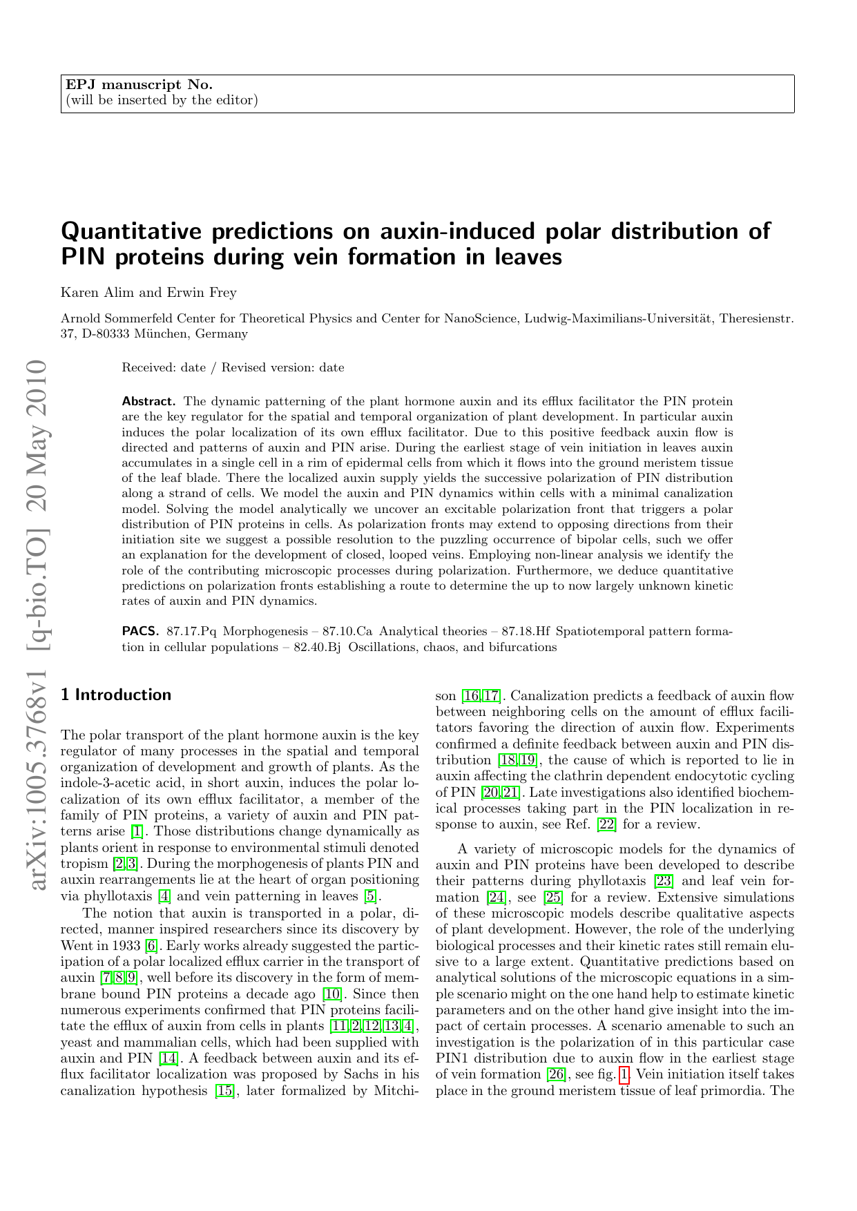
A variety of microscopic models for the dynamics of auxin and PIN proteins have been developed to describe their patterns during phyllotaxis [23] and leaf vein formation [24], see [25] for a review. Extensive simulations of these microscopic models describe qualitative aspects of plant development. However, the role of the underlying biological processes and their kinetic rates still remain elusive to a large extent. Quantitative predictions based on analytical solutions of the microscopic equations in a simple scenario might on the one hand help to estimate kinetic parameters and on the other hand give insight into the impact of certain processes. A scenario amenable to such an investigation is the polarization of in this particular case PIN1 distribution due to auxin flow in the earliest stage of vein formation [26], see fig. 1. Vein initiation itself takes place in the ground meristem tissue of leaf primordia. The (B) Polarization of PIN distribution in a strand of cell due to auxin inflow from the left, indicating details of auxin and PIN dynamics. The weak acid auxin accumulates in the interior of a plant cell due to a gradient in pH. In the inner cell the charged anion is trapped and can only be transported outwards by help of efflux facilitators in the form of PIN proteins. The auxin transport from cell to cell has an efficiency eA. Auxin synthesis sA and degradation dA takes place in the inner cell. PIN proteins cycle between the bulk and the cell membrane by basal attachment sP and detachment rates dP . In addition, positive auxin net flow is modeled to feed back on these rates increasing the PIN attachment by gP . Along the strand of cells the color shading indicates relative concentration of auxin and PIN on either membrane.
positions of vein initiation sites are determined by auxin accumulation in “convergence” points, which lie in a rim of epidermal cells around the ground meristem tissue [19,27,28,29]. These single cells with high auxin concentration polarize towards the ground meristem and locally transport their auxin into a cell in the ground meristem tissue. This localized inflow triggers the successive polarization of PIN distributions along a strand of cells starting from the cell with auxin inflow [19]. The strand of polarized cells finally extends up to a previously existing strand of polarized cells, building the pre-pattern for the vascular network. Starting from the petiole of the leaf primordium the polarized cells differentiate then into vascular cells [30]. In particular second order veins in Arabidopsis thaliana exhibit PIN polarization in opposite directions starting from a single bipolar cell, which lies in the ground meristem below the auxin convergence point in the epidermal layer [19]. This yet unresolved behavior gives rise to the formation of closed vein loops when both oppositely polarized strands connect to already formed veins.
A resolution on the origin of bipolar cells is postulated by examination of a minimal canalization model for the polarization of PIN distribution due to auxin supply in a one-dimensional strand of cells. Performing a nonlinear analysis of the model
…(Full text truncated)…
This content is AI-processed based on ArXiv data.