Microcalcifications in mammogram have been mainly targeted as a reliable earliest sign of breast cancer and their early detection is vital to improve its prognosis. Since their size is very small and may be easily overlooked by the examining radiologist, computer-based detection output can assist the radiologist to improve the diagnostic accuracy. In this paper, we have proposed an algorithm for detecting microcalcification in mammogram. The proposed microcalcification detection algorithm involves mammogram quality enhancement using multirresolution analysis based on the dyadic wavelet transform and microcalcification detection by fuzzy shell clustering. It may be possible to detect nodular components such as microcalcification accurately by introducing shape information. The effectiveness of the proposed algorithm for microcalcification detection is confirmed by experimental results.
Deep Dive into Detection of Microcalcification in Mammograms Using Wavelet Transform and Fuzzy Shell Clustering.
Microcalcifications in mammogram have been mainly targeted as a reliable earliest sign of breast cancer and their early detection is vital to improve its prognosis. Since their size is very small and may be easily overlooked by the examining radiologist, computer-based detection output can assist the radiologist to improve the diagnostic accuracy. In this paper, we have proposed an algorithm for detecting microcalcification in mammogram. The proposed microcalcification detection algorithm involves mammogram quality enhancement using multirresolution analysis based on the dyadic wavelet transform and microcalcification detection by fuzzy shell clustering. It may be possible to detect nodular components such as microcalcification accurately by introducing shape information. The effectiveness of the proposed algorithm for microcalcification detection is confirmed by experimental results.
Breast cancer is the second leading cause of cancer deaths for women and is found as one in eight women in the United States. It is a disease in which cells in the tissues of the breast become abnormal and divide without order or control. These abnormal cells form too much tissue and become a tumor. According to WHO report, nearly two million women are diagnosed with breast cancer every year worldwide. The disease can be treated if discovered so early enough. The effective detection of breast cancer in earlier stage increases the survival rate. The appropriate method for early detection of pre cancerous symptoms is screening mammography, which has to be conducted as a regular test for women.
Calcification clusters are an early sign of breast Cancer. Microcalcifications are quite very small bits of calcium deposits present inside the soft breast tissue. It shows up in clusters or in patterns (like circles or lines) associated with extra cell activity in breast region.
Microcalcifications appear in mammogram image as small localized granular points with high brightness. It is not easy to detect by naked eye because of its miniaturized dimension. However about 10%-40% of Microcalcification clusters are missed by radiologists due to its small size and nonpalpable [1], [2]. To avoid these problems, a New CAD (computer Aided diagnosis) system has to be developed to improve the diagnostic rate. By incorporating the expert knowledge of radiologists, the CAD system can be made to provide a clear insight about the disease and saves the society from breast cancer.
Many researchers have proposed the algorithms for Microcalcification detection based on discrete wavelet transform, which is a powerful tool for analyzing the image hierarchically on the basis of scale. Some researchers have developed a CAD system using fuzzy clustering, artificial neural network and genetic algorithm. R. N. Strickland et al. [3], H.Yoshida et al. [4] used a wavelet transform to detect microcalcification and the fuzzy logic was tried by N.Pandey et al. [5]. Anne Strauss et al. [6] presented an identification scheme based on watershed Processing. Valverde et al. [7] used a deformable-based model for Microcalcification detection. Some of these studies detect approximately 70% to 80% of correct calcification. Objective of CAD system is to reduce the false positives and consistency of radiologists in image interpretation [8]. Naturally Microcalcifications are nodular in structure, other tissue such as mammary ducts blood vessels are linear in structure [9]. The Fuzzy shell clustering algorithm (FSC) is best in identifying circular objects present in an image [10]. In the proposed method, wavelet has been combined with fuzzy shell clustering (FSC) algorithm in order to mark the Microcalcification region The rest of this paper is organized as follows. Section II presents microcalcification enhancement by using dyadic wavelet transform; the detection part using Fuzzy shell clustering in Section III, Results obtained on execution of algorithm are presented in section IV and Conclusion as last section. Microcalcifications appear as subtle and bright spots, whose size varies from 0.3mm to 1mm in the mammogram image. It is not easy to enhance the microcalcification regions since surrounding dense breast tissue makes the abnormality areas almost invisible. Microcalcifications are high frequency in nature. So it can be extracted by using high pass filtering. But conventional enhancement technique like unsharp masking, homomorphic filters and high boost filtering tends to change the characteristics of microcalcification. To overcome these limitations microcalcification regions can be enhanced by dyadic wavelet transform without modifying characteristics of microcalcification.
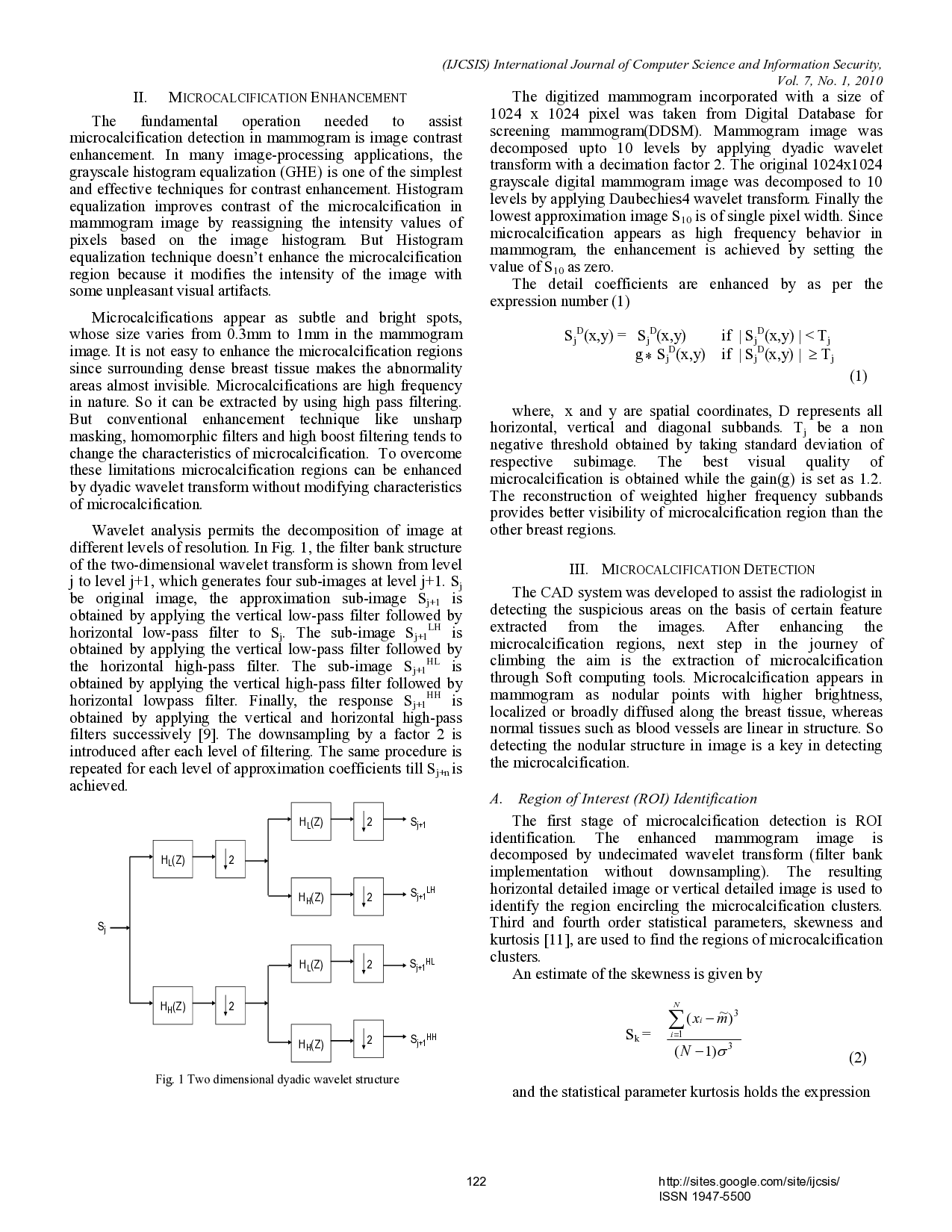
Wavelet analysis permits the decomposition of image at different levels of resolution. In Fig. 1, the filter bank structure of the two-dimensional wavelet transform is shown from level j to level j+1, which generates four sub-images at level j+1. S j be original image, the approximation sub-image S j+1 is obtained by applying the vertical low-pass filter followed by horizontal low-pass filter to S j . The sub-image S j+1
LH is obtained by applying the vertical low-pass filter followed by the horizontal high-pass filter. The sub-image S j+1 HL is obtained by applying the vertical high-pass filter followed by horizontal lowpass filter. Finally, the response S j+1 HH is obtained by applying the vertical and horizontal high-pass filters successively [9]. The downsampling by a factor 2 is introduced after each level of filtering. The same procedure is repeated for each level of approximation coefficients till S j+n is achieved. The digitized mammogram incorporated with a size of 1024 x 1024 pixel was taken from Digital Database for screening mammogram(DDSM). Mammogram image was decomposed upto 10 levels by applying dyadic wavelet transform with a decimation factor 2. The original 10
…(Full text truncated)…
This content is AI-processed based on ArXiv data.