Peeling and Sliding in Nucleosome Repositioning
We investigate the mechanisms of histone sliding and detachment with a stochastic model that couples thermally-induced, passive histone sliding with active motor-driven histone unwrapping. Analysis of a passive loop or twist defect-mediated histone sliding mechanism shows that diffusional sliding is enhanced as larger portions of the DNA is peeled off the histone. The mean times to histone detachment and the mean distance traveled by the motor complex prior to histone detachment are computed as functions of the intrinsic speed of the motor. Fast motors preferentially induce detachment over sliding. However, for a fixed motor speed, increasing the histone-DNA affinity (and thereby decreasing the passive sliding rate) increases the mean distance traveled by the motor.
💡 Research Summary
The paper presents a unified stochastic framework that couples two fundamentally different mechanisms governing nucleosome repositioning: passive, thermally driven sliding of the histone–DNA complex and active, motor‑driven peeling (unwrapping) of DNA from the histone core. The authors begin by reviewing the long‑standing debate over how nucleosomes move along DNA during transcription, replication, and repair. Historically, two separate models have been invoked. The first is a defect‑mediated sliding model, in which either a loop defect (a transient extra turn of DNA that propagates around the histone surface) or a twist defect (a localized over‑twist that diffuses) allows the histone octamer to shift one base pair at a time. The second is an active unwrapping model, wherein an ATP‑dependent motor (such as RNA polymerase, a helicase, or a remodeling complex) pulls DNA forward, sequentially breaking histone–DNA contacts and eventually ejecting the histone.
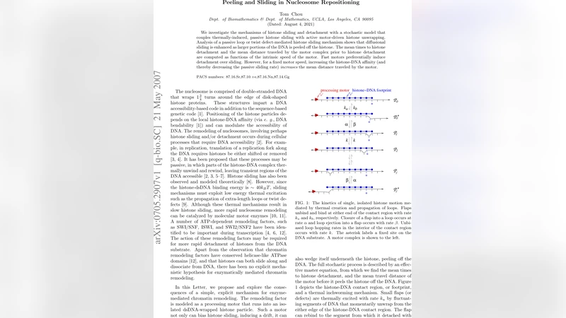
To capture the interplay of these processes, the authors construct a master equation that tracks the state of the nucleosome in terms of (i) the number of DNA base pairs still wrapped, (ii) the position of any sliding defect, and (iii) the progress of the motor along the DNA. The key kinetic parameters are:
* kₛ – the rate of creation (or propagation) of a loop/twist defect, which sets the effective diffusion coefficient D ≈ kₛ a² (a = 0.34 nm, the base‑pair spacing); * kₚ – the forward peeling rate, i.e., the probability per unit time that a wrapped base pair is released by the motor; * kᵣ – the reverse re‑wrapping rate, reflecting the intrinsic histone–DNA affinity; * v₀ – the intrinsic speed of the motor when it is not impeded by the nucleosome.
By solving the master equation analytically (and confirming with kinetic Monte‑Carlo simulations), the authors obtain closed‑form expressions for two observables of biological relevance:
- Mean detachment time ⟨τ⟩ – the average time required for the nucleosome to become completely unwrapped and released.
- Mean motor travel distance ⟨L⟩ – the average distance the motor travels along DNA before the nucleosome detaches.
The analysis reveals two distinct regimes. In the “fast‑motor” regime (v₀ ≫ kₛ a), the motor’s pulling force overwhelms the defect‑mediated diffusion. The nucleosome is ripped off almost immediately, so ⟨L⟩ ≈ v₀ ⟨τ⟩ and sliding contributes negligibly. In the opposite “slow‑motor” regime (v₀ ≲ kₛ a), the defect diffusion is sufficiently rapid that the nucleosome can slide many base pairs before the motor reaches the remaining contacts. Consequently, the histone travels a measurable distance along DNA, and the motor must traverse a longer stretch before detachment occurs.
A particularly insightful result concerns the role of histone–DNA affinity. Increasing affinity (e.g., by reducing kₚ or increasing kᵣ) lowers the passive sliding rate kₛ because the defect encounters a higher energetic barrier. Counter‑intuitively, this leads to a larger ⟨L⟩ for a fixed motor speed: the motor must “walk” farther because the nucleosome is less likely to be peeled off locally and instead slides away. This prediction aligns with experimental observations that nucleosomes containing post‑translationally modified histones (such as acetylated H3/H4, which weaken DNA binding) are more readily displaced by polymerases, whereas highly stable nucleosomes require the polymerase to travel further before successful eviction.
The authors map the phase space defined by (v₀, kₛ) and identify a transition line separating sliding‑dominant from peeling‑dominant behavior. They argue that cellular factors—such as the presence of chromatin remodelers, the degree of histone modification, or the local concentration of ATP—can shift the system across this boundary, thereby regulating gene expression dynamics.
In the discussion, the paper proposes experimental tests of the model. Single‑molecule FRET assays could monitor the distance between labeled DNA and histone cores while a motor protein is driven at controlled speeds, directly measuring the dependence of sliding diffusion on the amount of DNA peeled. Optical‑trap experiments could quantify the force–velocity relationship of a motor encountering a nucleosome and compare the observed detachment distances with the theoretical ⟨L⟩ curves. Moreover, microfluidic devices that allow simultaneous observation of multiple nucleosomes under varying ATP concentrations could validate the predicted scaling of ⟨τ⟩ with motor speed and histone affinity.
Overall, the study provides a quantitative bridge between two historically separate concepts of nucleosome mobility. By integrating passive diffusion and active unwrapping into a single stochastic description, it offers a versatile platform for exploring how transcription factors, remodelers, and histone modifications collectively shape chromatin dynamics. Future extensions could incorporate multiple motors, cooperative remodeling complexes, or the effect of DNA supercoiling, thereby moving closer to a comprehensive physical model of chromatin behavior in vivo.
Comments & Academic Discussion
Loading comments...
Leave a Comment