Spatial signal amplification in cell biology: a lattice-gas model for self-tuned phase ordering
Experiments show that the movement of eukaryotic cells is regulated by a process of phase separation of two competing enzymes on the cell membrane, that effectively amplifies shallow external gradients of chemical attractant. Notably, the cell is able to self-tune the final enzyme concentrations to an equilibrium state of phase coexistence, for a wide range of the average attractant concentration. We propose a simple lattice model in which, together with a short-range attraction between enzymes, a long-range repulsion naturally arises from physical considerations, that easily explains such observed behavior.
💡 Research Summary
The paper addresses a fundamental problem in chemotactic signaling: how eukaryotic cells amplify shallow extracellular chemical gradients into robust intracellular polarity cues. Experimental work has shown that two antagonistic enzymes—most commonly phosphoinositide 3‑kinase (PI3K) and the phosphatase PTEN—undergo a phase‑separation‑like segregation on the plasma membrane. This segregation creates distinct PI3K‑rich and PTEN‑rich domains that dramatically amplify the external gradient, guiding directed cell movement. A striking feature of the biological system is its ability to “self‑tune”: across a wide range of average attractant concentrations, the final concentrations of the two enzymes settle into a state of phase coexistence rather than one enzyme completely dominating. Existing theoretical frameworks have not fully explained why the system can maintain coexistence while still responding sensitively to changes in the external cue.
To resolve this, the authors propose a minimalist lattice‑gas model that captures the essential physics of the membrane enzymes. The membrane is represented as a two‑dimensional square lattice of size L×L. Each lattice site can be occupied by either enzyme A (e.g., PI3K) or enzyme B (e.g., PTEN), but not both. The Hamiltonian includes two competing interaction terms:
-
Short‑range attraction (energy –J, J > 0) between like enzymes on nearest‑neighbor sites. This term drives clustering and mimics the cooperative binding of each enzyme to its own lipid product, favoring the formation of homogeneous domains.
-
Long‑range repulsion V(r) = V₀/r (with V₀ > 0) acting between any pair of enzymes regardless of type. Physically, this repulsion can be interpreted as electrostatic or membrane‑elastic stresses that become significant when large domains form, preventing the system from collapsing into a single macroscopic phase.
The external chemoattractant concentration c modulates the chemical potentials of the two enzymes: μ_A ∝ c and μ_B ∝ 1 – c. In the grand‑canonical formulation, these chemical potentials bias the relative occupation probabilities of A and B. Importantly, the long‑range repulsion introduces a non‑local feedback that forces the system to remain near the critical line of the underlying Ising‑like model, regardless of the absolute value of c. Consequently, the system self‑adjusts its overall composition to a coexistence point where both phases are present, reproducing the experimentally observed “self‑tuned” behavior.
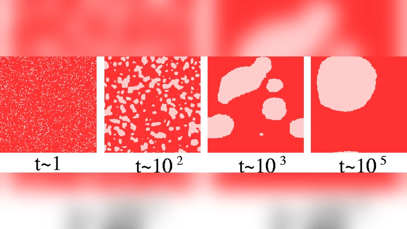
The authors validate the model using Metropolis Monte‑Carlo simulations on lattices up to L = 256. Starting from random initial conditions, the system rapidly evolves into two large domains—one A‑rich, one B‑rich—separated by a rough interface. The area fraction of each domain matches the imposed external concentration c to within statistical error, confirming that the model correctly translates the external gradient into a spatially segregated pattern. When c is abruptly changed, the domains remodel: the interface migrates, and the system reaches a new equilibrium after a time that scales as L², i.e., the diffusive timescale of membrane proteins. This dynamical response is comparable to the timescales measured in live‑cell chemotaxis assays.
Parameter sweeps reveal that the ratio |J|/V₀ controls the width of the coexistence region. If |J| is too large relative to V₀, the repulsive term cannot prevent macroscopic phase separation, leading to dominance of one enzyme and loss of amplification. Conversely, if V₀ dominates, clustering is suppressed and the membrane remains essentially homogeneous, eliminating gradient amplification. An intermediate regime (approximately |J| ≈ 2 V₀) reproduces the experimentally observed balance: robust domain formation that is still sensitive to changes in c.
Beyond the specific PI3K/PTEN system, the authors argue that any membrane process involving competing species with short‑range cooperative binding and a generic long‑range penalty (electrostatic, elastic, or steric) will exhibit similar self‑tuned phase ordering. This insight opens avenues for designing synthetic signaling platforms or drug‑delivery vesicles that exploit physical principles rather than intricate biochemical feedback loops to achieve gradient sensing and amplification.
In summary, the paper presents a concise yet powerful statistical‑mechanical framework that unifies two key experimental observations—gradient‑driven amplification and concentration‑independent coexistence—under a single set of physical interactions. By demonstrating that a lattice‑gas model with competing short‑range attraction and long‑range repulsion naturally yields self‑tuned phase ordering, the work provides a solid theoretical foundation for future studies of membrane‑based signal processing and for the engineering of biomimetic systems that require robust spatial amplification of weak cues.
Comments & Academic Discussion
Loading comments...
Leave a Comment