Mechanical deformation of monocytic THP-1 cells : occurrence of two seqential phases with differential sensitivity to metabolic inhibitors
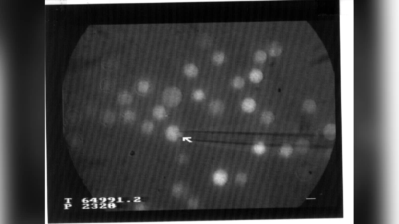
Blood leukocytes can exhibit extensive morphological changes during their passage through small capillary vessels. The human monocytic THP-1 cell line was used to explore the metabolic dependence of these shape changes. Cells were aspirated into micropipettes for determination of the rate of protrusion formation. They were then released and the kinetics of morphological recovery was studied. Results were consistent with Evans’ model (Blood, 64 : 1028, 1984) of a viscous liquid droplet surrounded by a tensile membrane. The estimated values of cytoplasmic viscosity and membrane tension were 162 Pa.s and 0.0142 millinewton/m respectively. The influence of metabolic inhibitors on cell mechanical behaviour was then studied : results strongly suggested that deformation involved two sequential phases. The cell elongation rate measured during the first 30 seconds following the onset of aspiration was unaffected by azide, an inhibitor of energy production, and it was about doubled by cytochalasin D, a microfilament inhibitor, and colchicine, a microtubule inhibitor. However, during the following two minutes, deformation was almost abolished in cells treated with azide and cytochalasin D, whereas the protrusion of control cells exhibited about threefold length increase. It is concluded that, although cells seemed to deform as passive objects, active metabolic processes were required to allow extensive morphological changes triggered by external forces.
💡 Research Summary
The study investigates how human monocytic THP‑1 cells deform under mechanical stress and how metabolic and cytoskeletal inhibitors influence this process. Using a micropipette aspiration technique, the authors measured the rate at which a protrusion (or “tongue”) forms when a cell is drawn into the pipette, and then recorded the kinetics of shape recovery after the cell is released. The deformation curves fit Evans’ classic model of a viscous liquid droplet bounded by a tensile membrane, allowing the authors to estimate a cytoplasmic viscosity of 162 Pa·s and a membrane tension of 0.0142 mN·m⁻¹. These values support the notion that, at a first approximation, a cell behaves as a passive visco‑elastic object.
To probe the role of active processes, the authors applied three pharmacological agents: sodium azide (an inhibitor of oxidative phosphorylation, thus blocking ATP production), cytochalasin D (which disrupts actin microfilaments), and colchicine (which depolymerises microtubules). The experiments revealed two temporally distinct phases of deformation.
Phase 1 (0–30 s after aspiration onset). In this early window, azide had virtually no effect on the rate of protrusion formation, indicating that ATP‑driven processes are not required for the initial, rapid deformation. By contrast, both cytochalasin D and colchicine doubled the elongation rate. This suggests that the intact actin cortex and microtubule network normally provide a mechanical brake; when these structures are compromised, the external suction more efficiently pulls the cell into the pipette.
Phase 2 (30 s–2 min). In the later interval, the picture reverses. Cells treated with azide alone, or with cytochalasin D, exhibited almost complete abolition of further elongation, whereas control cells continued to extend, achieving roughly a three‑fold increase in protrusion length. The combination of azide and cytochalasin D was especially potent in suppressing deformation. Colchicine alone did not markedly affect this later phase, implying that microtubules are less critical for sustained shape change. The data therefore indicate that the prolonged, extensive deformation requires active, ATP‑dependent remodeling of the actin cytoskeleton.
The authors conclude that, although the initial response to an external force can be described by passive fluid‑membrane mechanics, the ability of a leukocyte to undergo large, sustained morphological changes depends on active metabolic processes. In physiological terms, this two‑stage behavior may reflect how circulating leukocytes negotiate narrow capillaries: a rapid, passive “squeezing” followed by an energy‑requiring, actin‑driven remodeling that permits continued elongation, adhesion, or transmigration.
Methodologically, the work demonstrates the value of combining quantitative micropipette aspiration with targeted pharmacology to dissect the mechanical contributions of viscosity, membrane tension, and cytoskeletal dynamics. The derived physical parameters (162 Pa·s viscosity, 0.0142 mN·m⁻¹ tension) are consistent with earlier reports for leukocytes, reinforcing the validity of the Evans model for these cells.
From a broader perspective, the findings highlight that purely mechanical models are insufficient to predict leukocyte behavior under prolonged mechanical stress. Incorporating ATP‑dependent actin turnover, cortical tension regulation, and possibly signaling pathways that couple metabolic state to cytoskeletal remodeling will be essential for realistic simulations of immune cell trafficking, inflammation, and related pathologies. Moreover, the differential sensitivity to azide versus cytoskeletal drugs suggests potential therapeutic windows: targeting metabolic pathways could blunt excessive leukocyte deformation and extravasation in inflammatory diseases, whereas modulating actin dynamics might affect the early mechanical “squeezing” phase.
In summary, the paper provides compelling evidence that THP‑1 cell deformation proceeds through a fast, passive phase governed mainly by existing structural resistance, followed by a slower, active phase that is critically dependent on ATP production and actin filament dynamics. This dual‑phase model refines our understanding of leukocyte mechanics and underscores the necessity of integrating biophysical and biochemical viewpoints when studying cell deformation in health and disease.
Comments & Academic Discussion
Loading comments...
Leave a Comment