A computer-aided detection (CAD) system for the identification of pulmonary nodules in low-dose multi-detector computed-tomography (CT) images has been developed in the framework of the MAGIC-5 Italian project. One of the main goals of this project is to build a distributed database of lung CT scans in order to enable automated image analysis through a data and cpu GRID infrastructure. The basic modules of our lung-CAD system, consisting in a 3D dot-enhancement filter for nodule detection and a neural classifier for false-positive finding reduction, are described. The system was designed and tested for both internal and sub-pleural nodules. The database used in this study consists of 17 low-dose CT scans reconstructed with thin slice thickness (~300 slices/scan). The preliminary results are shown in terms of the FROC analysis reporting a good sensitivity (85% range) for both internal and sub-pleural nodules at an acceptable level of false positive findings (1-9 FP/scan); the sensitivity value remains very high (75% range) even at 1-6 FP/scan
Deep Dive into Automated detection of lung nodules in low-dose computed tomography.
A computer-aided detection (CAD) system for the identification of pulmonary nodules in low-dose multi-detector computed-tomography (CT) images has been developed in the framework of the MAGIC-5 Italian project. One of the main goals of this project is to build a distributed database of lung CT scans in order to enable automated image analysis through a data and cpu GRID infrastructure. The basic modules of our lung-CAD system, consisting in a 3D dot-enhancement filter for nodule detection and a neural classifier for false-positive finding reduction, are described. The system was designed and tested for both internal and sub-pleural nodules. The database used in this study consists of 17 low-dose CT scans reconstructed with thin slice thickness (~300 slices/scan). The preliminary results are shown in terms of the FROC analysis reporting a good sensitivity (85% range) for both internal and sub-pleural nodules at an acceptable level of false positive findings (1-9 FP/scan); the sensitivit
Automated detection of lung nodules in low-dose computed tomography
D. Cascioa, S.C. Cheranb,c, A. Chincarinid, G. De Nunzioe, P. Deloguf,g, M.E. Fantaccif,g,
G. Garganoh,i, I. Gorig, G. L. Masalal,m, A. Preite Martinezn, A. Reticog, M. Santoroo,
C. Spinellip, T. Tarantinoq
aDipartimento di Fisica e Tecnologie Relative, Università di Palermo, Italy
bDipartimento di Fisica, Università di Genova, Italy
cIstituto Nazionale di Fisica Nucleare, Sezione di Torino, Italy
dIstituto Nazionale di Fisica Nucleare, Sezione di Genova, Italy
eDipartimento di Scienza dei Materiali, Università di Lecce, Italy
fDipartimento di Fisica, Università di Pisa, Italy
gIstituto Nazionale di Fisica Nucleare, Sezione di Pisa, Italy
hDipartimento Interateneo di Fisica M. Merlin, Università di Bari, Italy
iIstituto Nazionale di Fisica Nucleare, Sezione di Bari, Italy
lStruttura Dipartimentale di Matematica e Fisica, Università di Sassari, Italy
mIstituto Nazionale di Fisica Nucleare, Sezione di Cagliari, Italy
nCentro Studi e Ricerche Enrico Fermi, Roma, Italy
oDipartimento di Scienze Fisiche, Università di Napoli, Italy
pUnità Operativa Radiodiagnostica 2, Azienda Ospedaliera Universitaria Pisana, Pisa, Italy
qDivisione di Radiologia Diagnostica e Interventistica del Dipartimento di Oncologia, Trapianti e Nuove
Tecnologie in Medicina, Università di Pisa, Italy
Abstract. A computer-aided detection (CAD) system for the identification of
pulmonary nodules in low-dose multi-detector computed-tomography (CT) images has
been developed in the framework of the MAGIC-5 Italian project. One of the main
goals of this project is to build a distributed database of lung CT scans in order to enable
automated image analysis through a data and cpu GRID infrastructure.
The basic modules of our lung-CAD system, consisting in a 3D dot-enhancement filter
for nodule detection and a neural classifier for false-positive finding reduction, are
described. The system was designed and tested for both internal and sub-pleural
nodules. The database used in this study consists of 17 low-dose CT scans reconstructed
with thin slice thickness (~300 slices/scan). The preliminary results are shown in terms
of the FROC analysis reporting a good sensitivity (85% range) for both internal and
sub-pleural nodules at an acceptable level of false positive findings (1–9 FP/scan); the
sensitivity value remains very high (75% range) even at 1–6 FP/scan.
Keywords: Computer-aided detection (CAD); low-dose computed tomography (LDCT); thin-slice CT;
lung cancer screening.
- Introduction
One of the early markers of lung cancer is the presence of non-calcified small
pulmonary nodules. Their radiological appearance shows pseudo-spherical objects
usually characterized by low contrast and CT values similar to those of blood vessels
and airway walls to which they can be connected. Computed Tomography (CT) has
been shown as the most sensitive imaging modality for the detection of small
pulmonary nodules, particularly since the introduction of the multi-detector-row CT
technology [1]. Clinical programs based on low-dose CT (LDCT) are regarded as
promising screening techniques for early-stage lung cancers [2]. However, the amount
of data to be interpreted for each patient can be very large due to the thin sections
usually reconstructed in these protocols.
To support radiologists in the identification of early-stage pathological objects,
researchers have begun over one decade ago to explore computer-aided detection
(CAD) methods in this indication.
The First Italian Randomized Controlled Trial (ITALUNG-CT) that aims to study the
potential impact of screening on a high-risk population using low-dose helical CT with
thin collimation was recently started [3].
In the framework of MAGIC-5 collaboration funded by Istituto Nazionale di Fisica
Nucleare (INFN) and Ministero dell’Università e della Ricerca (MIUR), we have
developed a CAD system for pulmonary nodule identification, based on the analysis of
images acquired from the Pisa centre of the ITALUNG-CT trial. The system was
designed and tested for both internal and sub-pleural lung nodules. - The CAD strategy
The database used in this study consists of 17 low-dose CT scans acquired with a 4
slices spiral CT scanner according to a low-dose protocol (screening setting: 140 kV, 20
mA), with a 1.25 mm slice collimation [3]. The average number of slices per scan is
about 300 with 512×512 pixel matrix, a pixel size ranging from 0.53 to 0.74 mm and 12
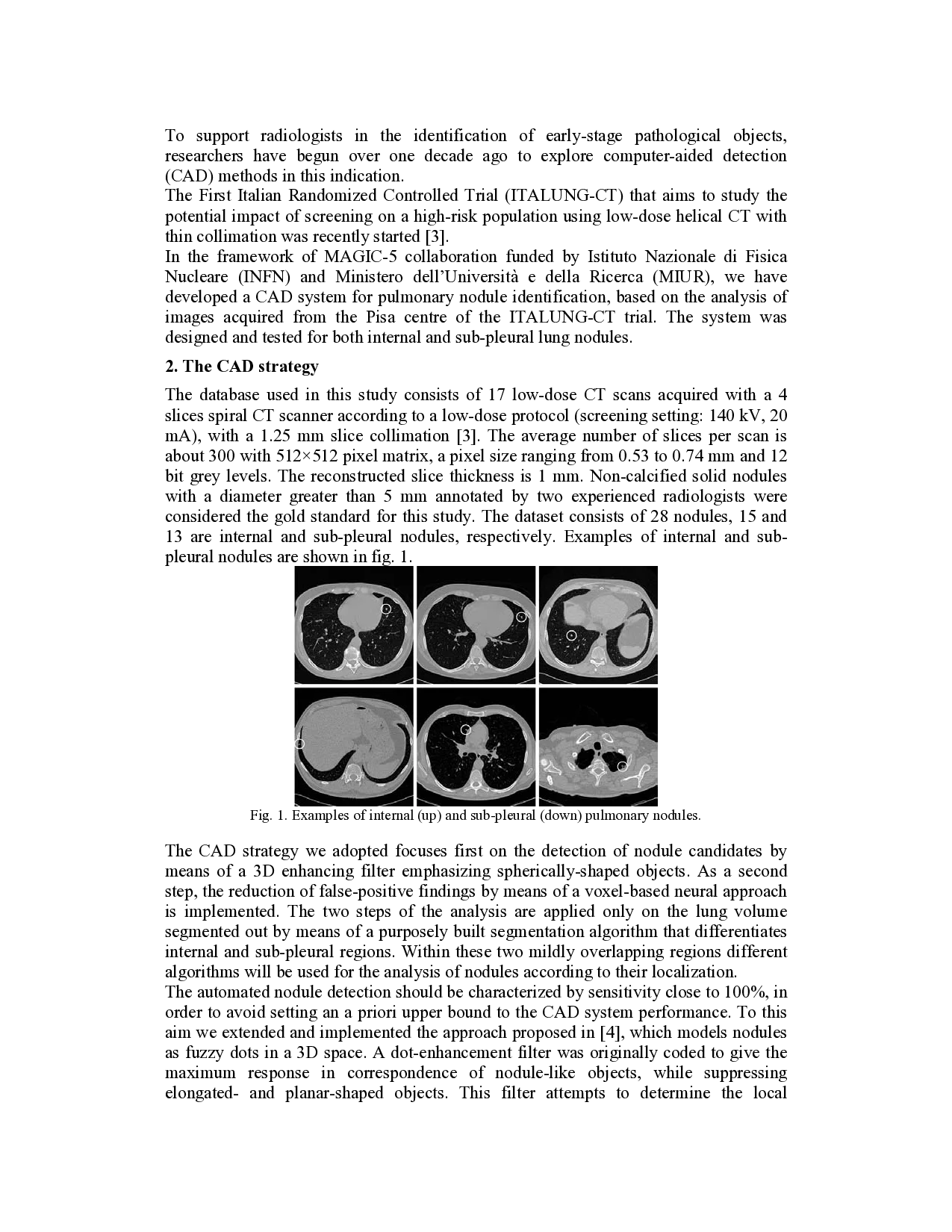
bit grey levels. The reconstructed slice thickness is 1 mm. Non-calcified solid nodules
with a diameter greater than 5 mm annotated by two experienced radiologists were
considered the gold standard for this study. The dataset consists of 28 nodules, 15 and
13 are internal and sub-pleural nodules, respectively. Examples of internal and sub-
pleural nodules are shown in fig. 1.
Fig. 1. Examples of internal (up) and sub-ple
…(Full text truncated)…
This content is AI-processed based on ArXiv data.