Silver staining of proteins in polyacrylamide gels
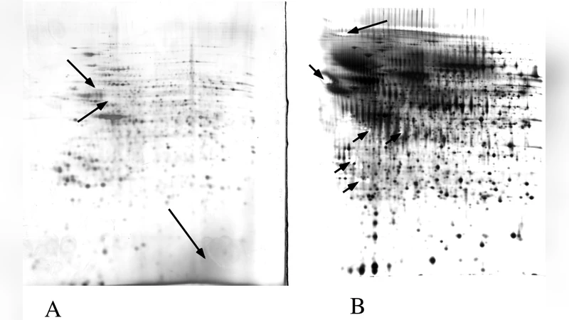
Silver staining is used to detect proteins after electrophoretic separation on polyacrylamide gels. It combines excellent sensitivity (in the low nanogram range) with the use of very simple and cheap equipment and chemicals. It is compatible with downstream processing, such as mass spectrometry analysis after protein digestion. The sequential phases of silver staining are protein fixation, then sensitization, then silver impregnation and finally image development. Several variants of silver staining are described here, which can be completed in a time range from 2 h to 1 d after the end of the electrophoretic separation. Once completed, the stain is stable for several weeks.
💡 Research Summary
The paper provides a comprehensive guide to silver staining of proteins separated by polyacrylamide gel electrophoresis, emphasizing its exceptional sensitivity, low cost, and compatibility with downstream analyses such as mass spectrometry. The method is divided into four sequential phases: fixation, sensitization, silver impregnation, and development.
In the fixation step, proteins are immobilized within the gel matrix using a mixture of methanol, acetonitrile, and formic acid (typically 50 % methanol, 10 % acetonitrile, 5 % formic acid). This prevents protein diffusion during subsequent steps and preserves the electrophoretic pattern. Alternative fixation with glutaraldehyde‑formaldehyde improves structural preservation for delicate samples.
Sensitization follows fixation and enhances the affinity of proteins for silver ions. Commonly, a 0.02 % sodium thiosulfate solution is applied, sometimes supplemented with a small amount of formic acid to adjust the pH to 5.5–6.0. The sensitization period is brief (5–15 min); over‑sensitization leads to increased background staining.
Silver impregnation introduces silver ions into the gel by soaking it in a 0.1 % silver nitrate solution for 20–30 min at room temperature (20–25 °C). The previously sensitized protein sites bind silver ions preferentially, while excess ions are removed by thorough rinsing.
Development is the final visualisation step. A developer containing 0.1 % formic acid, 0.05 % ascorbic acid, and 0.05 % sodium bicarbonate reduces bound silver ions to metallic silver, producing visible bands. Development is rapid (1–5 min) and must be stopped promptly by rinsing to avoid over‑development and high background. Temperature and pH control are critical for reproducibility.
The authors describe several protocol variations that trade off speed, background, and sensitivity. Low‑temperature development (4 °C) combined with sodium acetate buffers reduces background dramatically and pushes detection limits to ~0.5 ng of protein. A glutaraldehyde‑formaldehyde fixation‑sensitization scheme further improves signal‑to‑noise for low‑abundance proteins.
Compatibility with mass spectrometry is a central concern. Silver particles can suppress peptide ionisation, but the paper demonstrates that post‑staining treatments—such as a brief wash in a formic‑citrate solution or direct in‑gel trypsin digestion—effectively remove or bypass silver interference, allowing reliable MALDI‑TOF or LC‑MS/MS analysis.
Time efficiency is highlighted: a rapid “standard” protocol can be completed within two hours after electrophoresis, while more sensitive, low‑background variants may require 12–24 hours due to additional sensitisation and development steps. Regardless of the chosen variant, the stained gels remain stable for several weeks at room temperature, making them suitable for archival storage and high‑quality image acquisition.
Overall, the paper positions silver staining as a versatile, high‑sensitivity detection method that balances cost, simplicity, and downstream analytical compatibility. It provides detailed parameters for each step, discusses the chemical rationale behind them, and offers practical solutions to known limitations such as mass‑spectrometric suppression. This makes the work a valuable reference for both newcomers and experienced proteomics laboratories seeking to implement or optimise silver staining in their workflows.
Comments & Academic Discussion
Loading comments...
Leave a Comment