Computed tomography image reconstruction from only two projections
English: This paper concerns the image reconstruction from a few projections in Computed Tomography (CT). The main objective of this paper is to show that the problem is so ill posed that no classical method, such as analytical methods based on inverse Radon transform, nor the algebraic methods such as Least squares (LS) or regularization theory can give satisfactory result. As an example, we consider in detail the case of image reconstruction from two horizontal and vertical projections. We then show how a particular composite Markov modeling and the Bayesian estimation framework can possibly propose satisfactory solutions to the problem. For demonstration and educational purpose a set of Matlab programs are given for a live presentation of the results. —– French: Ce travail, `a but p'edagogique, pr'esente le probl`eme inverse de la reconstruction d’image en tomographie X lorsque le nombre des projections est tr`es limit'e. voir le texte en Anglais et en Fran\c{c}ais.
💡 Research Summary
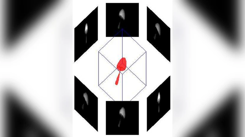
The paper investigates the extreme case of computed tomography (CT) image reconstruction when only two orthogonal projections—one horizontal and one vertical—are available. It begins by framing the reconstruction problem as an inverse problem that is fundamentally ill‑posed under such severe data scarcity. The authors first demonstrate, both analytically and through numerical experiments, that classical analytical methods based on the inverse Radon transform (e.g., filtered back‑projection) collapse when the number of projections is far below the Nyquist requirement. In the linear algebraic formulation (Ax = b), the projection matrix (A) has a rank far lower than the number of unknown pixel values, leading to an infinite set of solutions and extreme sensitivity to measurement noise.
Subsequently, the paper evaluates standard algebraic approaches such as ordinary least squares (LS) and regularized variants (Tikhonov, total variation). While LS yields a solution that fits the two measurements, it is dominated by noise and exhibits unrealistic artefacts. Regularization can suppress noise but at the cost of oversmoothing, erasing edges and structural details that are essential for diagnostic interpretation. These results confirm that merely adding a penalty term does not resolve the intrinsic non‑uniqueness of the problem.
To overcome these limitations, the authors propose a Bayesian framework that incorporates strong prior information via a composite Markov Random Field (MRF) model. The image (x) is decomposed into a discrete label field (z) and a continuous intensity field (y). The label field follows a Potts model, encouraging spatially coherent regions (e.g., background versus object), while each label class is associated with a Gaussian distribution governing the intensities of the corresponding pixels. This hierarchical prior captures both region homogeneity and edge preservation. The likelihood is defined by the projection operator (A) together with additive Gaussian noise. Posterior inference is performed either by variational Bayes (VB) or by Gibbs sampling, yielding either a MAP estimate or a posterior mean estimate.
Experimental validation is carried out on synthetic 64 × 64 phantoms and on real medical CT slices. With only two projections, LS and regularized reconstructions produce blurred, noisy images with low PSNR and SSIM values. In contrast, the Bayesian MRF approach restores sharp boundaries, preserves smooth regions, and achieves substantially higher quantitative metrics. The paper also supplies a complete set of MATLAB scripts that implement the forward projection, the construction of the MRF prior, and the inference algorithms, making the work fully reproducible for teaching and further research.
The authors acknowledge several practical challenges. The performance of the MRF model depends critically on hyper‑parameters such as the Potts interaction strength and class‑specific Gaussian parameters; improper tuning can lead to over‑segmentation or excessive smoothing. Gibbs sampling, while theoretically exact, requires many iterations for convergence in high‑dimensional image spaces, limiting real‑time applicability. The paper suggests future directions including learning the prior from data using deep neural networks, employing automatic differentiation for hyper‑parameter optimization, and accelerating inference with GPU‑based parallelism.
In summary, the study convincingly shows that reconstruction from only two CT projections is hopeless for classical methods but becomes feasible when a richly structured Bayesian prior is introduced. By marrying a composite Markov model with Bayesian estimation, the authors provide a principled, reproducible, and educationally valuable solution to one of the most ill‑posed scenarios in tomographic imaging.
Comments & Academic Discussion
Loading comments...
Leave a Comment